Question Of The Day #50

question of the day

Which of the following is the most appropriate next step in management for this patient’s condition?

This patient presents to the Emergency Department with altered mental status.  This presenting symptom can be due to a large variety of etiologies, including hypoglycemia, sepsis, toxic ingestions, electrolyte abnormalities, stroke, and more.  The management and evaluation of a patient with altered mental status depends on the primary assessment of the patient (“ABCs”, or Airway, Breathing, Circulation) to identify any acute life-threatening conditions that need to be managed emergently, the history, and the physical examination.  One mnemonic that may help in remembering the many causes of altered mental status is “AEIOUTIPS”.  The table below outlines this mnemonic.

ALTERED MENTAL STATUS

This patient arrives hyperthermic, tachycardic in atrial fibrillation, diaphoretic, and altered with psychotic behavior.  Thyroid storm, the most severe manifestation of hyperthyroidism, should always be on the differential diagnosis in patients with fever and altered mental status.  Other considerations are sepsis, sympathomimetic overdose, anticholinergic overdose, serotonin syndrome, and pheochromocytoma. 

This patient has thyroid storm, a life-threatening endocrine emergency that requires prompt recognition and treatment.  Symptoms of thyroid storm include altered mental status, psychosis, seizures, coma, tachycardia, atrial fibrillation, high-output heart failure, dyspnea, vomiting, diarrhea, weight loss, and anterior neck enlargement.  Severe hyperthyroidism should have a low-undetectable TSH level with elevated T3/T4 levels, but in acute illness these levels may be unreliable.  For this reason, the diagnosis and treatment of thyroid storm should be based on clinical grounds.

An anticholinergic toxidrome can appear similar to this patient with tachycardia, hypertension, agitation, and altered mental status.  A key differentiating factor is diaphoresis.  Patients with anticholinergic ingestions should have dry skin, not wet skin. The treatment for anticholinergic toxicity is benzodiazepines and IV physostigmine (Choice A) if symptoms are unresponsive to benzodiazepines.  Physostigmine is not the best next step in this scenario. 

Treatment of thyroid storm is algorithmic.  First, beta blockade (Choice C) should be given to control the heart rate and block T4 to T3 conversion, next anti-thyroid medications (Methimazole or Propylthiouracil (Choice D)) should be given to block thyroid hormone synthesis, and lastly corticosteroids and inorganic iodine (Choice B) can be given to block release of stored thyroid hormone.  The best next step in managing this patient with thyroid storm is administration of IV Propranolol (Choice C).  Propranolol helps manage the tachycardia, systemic symptoms, and also inhibits conversion of T4 to T3. 

 Correct Answer: C

References

Cite this article as: Joseph Ciano, USA, "Question Of The Day #50," in International Emergency Medicine Education Project, August 13, 2021, https://iem-student.org/2021/08/13/question-of-the-day-50/, date accessed: April 27, 2024

Question Of The Day #49

question of the day
Which of the following is the most likely cause of this patient’s condition?

This patient presents to the Emergency Department with altered mental status.  This presenting symptom can be due to a large variety of etiologies, including hypoglycemia, sepsis, toxic ingestions, electrolyte abnormalities, stroke, and more.  The management and evaluation of a patient with altered mental status depends on the primary assessment of the patient (“ABCs”, or Airway, Breathing, Circulation) to identify any acute life-threatening conditions that need to be managed emergently, the history, and the physical examination.  One mnemonic that may help in remembering the many causes of altered mental status is “AEIOUTIPS”.  The table below outlines this mnemonic.

ALTERED MENTAL STATUS

Hyperthermia (or hypothermia) can cause altered mental status.  This patient arrives with altered mental status, severe hyperthermia, tachycardia, tachypnea, and hypotension.  The history of the patient running outside for exercise should raise concern for hyperthermia related to excess heat production due to overexertion.  This should narrow the differential diagnoses to heat exhaustion (Choice B) and heat stroke (Choice C).  Both heat exhaustion and heat stroke are marked by hyperthermia with temperatures often over 40ᵒC. Additional symptoms include weakness, nausea, vomiting, myalgias, syncope, and headache.  The differentiating factor between heat exhaustion and heat stroke is altered mental status and sweating.  Patients with heat exhaustion lack altered mental status and should still be able to thermoregulate through sweating.  On the contrary, heat stroke patients are more severely ill as they have altered mental status and can no longer thermoregulate with sweating.  The treatment in both conditions should be early and aggressive cooling measures.  This includes full body immersion in an ice bath, removal of clothes, and cold IV fluids.  Internal cooling with gastric, bladder, pleural, or peritoneal lavage with cold fluids can be done on more sick patients.  Antipyretic medications, like NSAIDs and paracetamol, have no benefit in patients with severe hyperthermia.  Evaluation for rhabdomyolysis, kidney failure, liver failure, sepsis, or other organ dysfunction should also be a part of the evaluation of hyperthermic patients.

Sympathomimetic toxicity (Choice A) is possible, but less likely as the skin is dry and the history of exercise outdoors.  Sympathomimetic toxicity manifests as diaphoresis, tachycardia, hypertension, hyperthermia, and sometimes altered mental status.  Thyroid storm (Choice D) is another possibility.  This diagnosis can also present with similar vital signs, hyperthermia, and altered mental status.  Again, the history of outdoor exercise should point more towards heat exhaustion vs heat stroke.

The diagnosis of this patient is heat stroke (Choice C) as he has altered mental status and lacks wet skin.

Correct Answer: C

References

Cite this article as: Joseph Ciano, USA, "Question Of The Day #49," in International Emergency Medicine Education Project, August 6, 2021, https://iem-student.org/2021/08/06/question-of-the-day-49/, date accessed: April 27, 2024

Question Of The Day #48

question of the day

Which of the following is the most likely cause of this patient’s condition?

This patient presents to the Emergency Department with altered mental status.  This presenting symptom can be due to a large variety of etiologies, including hypoglycemia, sepsis, toxic ingestions, electrolyte abnormalities, stroke, and more.  The management and evaluation of a patient with altered mental status depends on the primary assessment of the patient (“ABCs”, or Airway, Breathing, Circulation) to identify any acute life-threatening conditions that need to be managed emergently, the history, and the physical examination.  One mnemonic that may help in remembering the many causes of altered mental status is “AEIOUTIPS”.  The table below outlines this mnemonic.

ALTERED MENTAL STATUS

The first step in managing this patient should be to treat the hypoxia with supplemental oxygen.  Prolonged hypoxia is dangerous and if left untreated, can cause brain damage.  Hypoxia can cause altered mental status, however, when this patient’s hypoxia is resolved, she remains somnolent and altered.  This should raise concern over an alternative etiology for the patient’s condition.      

The arterial blood gas demonstrates a low pH (acidosis), normal paO2, elevated paCO2 (hypercarbia), and a normal HCO3 (no metabolic compensation for acidosis).  The final interpretation of the ABG would be an acute respiratory acidosis without metabolic compensation.  Acute elevations of pCO2 can manifest as somnolence and altered mental status as seen in this patient.  This is known as hypercarbic or hypercapnic respiratory failure (Choice A).  This condition is caused by the inability to exhale CO2.  Risk factors include obstructive lung diseases (i.e., COPD), obesity, and obstructive sleep apnea.  Treatment involves treatment of hypoxia with supplemental oxygen, non-invasive positive pressure ventilation (i.e., BIPAP, CPAP, High Flow Nasal Cannula), and treatment of the underlying cause.

The patient’s arterial blood gas does not show hypoxic respiratory failure (Choice B).  Since treatment of the patient’s hypoxia does not improve the patient’s mental status, hypercarbic respiratory failure is more likely the underlying cause of the patient’s condition.  Opioid overdose (Choice C) can cause a similar ABG and patient presentation.  The normal size pupils and absent history of drug abuse makes this diagnosis less likely. Sepsis (Choice D) can trigger changes in mental status and cause respiratory failure, however, the absence of infectious symptoms and the presence of obesity and COPD support hypercarbic respiratory failure as the more likely underlying cause. 

Correct Answer: A

References

Cite this article as: Joseph Ciano, USA, "Question Of The Day #48," in International Emergency Medicine Education Project, July 30, 2021, https://iem-student.org/2021/07/30/question-of-the-day-48/, date accessed: April 27, 2024

Question Of The Day #47

question of the day

Which of the following is the most appropriate next step in management for this patient’s condition?

This patient presents to the Emergency Department with altered mental status.  This presenting symptom can be due to a large variety of etiologies, including hypoglycemia, sepsis, toxic ingestions, electrolyte abnormalities, stroke, and more.  The management and evaluation of a patient with altered mental status depends on the primary assessment of the patient (“ABCs”, or Airway, Breathing, Circulation) to identify any acute life-threatening conditions that need to be managed emergently, the history, and the physical examination.  One mnemonic that may help in remembering the many causes of altered mental status is “AEIOUTIPS”.  The table below outlines this mnemonic.

ALTERED MENTAL STATUS

The initial approach to all Emergency Department patients, especially those with abnormal vital signs, should include a primary survey (“ABCs”, or Airway, Breathing, Circulation).  This patient is breathing independently but at a significantly reduced rate and is hypoxic.  Hypoxia should prompt the administration of supplemental oxygen to the patient and reassessment of the SpO2.  The patient’s reduced respiratory rate, lethargy, and bilateral miosis (constricted pupils) should strongly hint at the possibility of opioid overdose.  Although the patient is lethargic and hypoxic, establishing a definitive airway (endotracheal intubation) should be avoided until after the antidote to opioid overdose is administered.  Naloxone is a mu-opioid receptor antagonist and functions as the antidote to opioid overdose.


Administration of 1000mL of 0.9% NaCl (Choice A) is unlikely to fix the patient’s clinical condition.  The patient needs naloxone to improve respiratory status.  25g of IV dextrose (Choice B) would be helpful if this patient’s altered mental status was from hypoglycemia.  A normal glucose level is provided in the question stem.  100mg of IV thiamine (Choice D) may be helpful in the case of Wernicke-Korsakoff Syndrome, a state of thiamine deficiency often associated with malnutrition and alcohol abuse.  Wernicke-Korsakoff Syndrome presents with vision disturbances, ataxia, and confusion.  Typically, this syndrome does not present with severe lethargy or depressed mental status as is seen in this patient.

The best next step in management is 1mg of IV naloxone (Choice C).  If given appropriately, naloxone can prevent the need for intubation.  Naloxone has a very short onset to action (~1min).  If suspicion for opioid overdose is high and there is an inadequate respiratory response after a single naloxone dose, repeat doses of naloxone are appropriate.  Naloxone can be administered in repeat boluses every 3-minutes to a total dose of 10mg IV.  Patients who respond appropriately to naloxone should be observed for recurrent respiratory depression as naloxone is cleared.  Need for repeat doses of naloxone indicates the need for a continuous naloxone infusion and hospital admission.  The typical infusion dose is 2/3 the “wake-up” dose given over 1 hour as a continuous infusion.  For example, if the patient responded to 1mg IV initially, the continuous infusion dose would be 0.6mg/hour of IV naloxone.

Correct Answer: C

References

Cite this article as: Joseph Ciano, USA, "Question Of The Day #47," in International Emergency Medicine Education Project, July 23, 2021, https://iem-student.org/2021/07/23/question-of-the-day-47/, date accessed: April 27, 2024

Question Of The Day #46

question of the day

Which of the following is the most appropriate next step in management for this patient’s condition?

This patient presents to the Emergency Department with altered mental status.  This presenting symptom can be due to a large variety of etiologies, including hypoglycemia, sepsis, toxic ingestions, electrolyte abnormalities, stroke, and more.  The management and evaluation of a patient with altered mental status depends on the primary assessment of the patient (“ABCs”, or Airway, Breathing, Circulation) to identify any acute life-threatening conditions that need to be managed emergently, the history, and the physical examination.  One mnemonic that may help in remembering the many causes of altered mental status is “AEIOUTIPS”.  The table below outlines this mnemonic.

ALTERED MENTAL STATUS

The serum chemistry results provided show elevated BUN and Creatinine with a BUN/Cr ratio of 21.3.  A BUN/Cr ratio greater than 20 indicates decreased perfusion to the kidneys, also known as pre-renal azotemia, which can indicate dehydration, hypovolemia, or shock.  The serum chemistry also shows a severely low sodium level.  Hyponatremia can present with a variety of symptoms, including weakness, fatigue, myalgias, nausea, vomiting, headaches, altered mental status, focal neurologic deficits, seizures, or coma.  Hyponatremia can be acute or chronic, asymptomatic or symptomatic, and mild or severe.  Sodium levels below 120 mEq/L are severely low.  Neurologic symptoms, such as seizures, altered mental status, and focal neurologic deficits, are also considered severe.  Treatment should be based on patient symptoms, rather than the sodium level, as it can be difficult to assess how acute or chronic the hyponatremia state is on initial evaluation.  The presence of any severe neurologic symptoms as is seen in this scenario should prompt administration of hypertonic saline (3% NaCl).  This allows for rapid correction of serum sodium levels, which should in turn relieve the neurologic symptoms.  A 100-150mL IV bolus of 3% NaCl can be given a second time if symptoms continue after 5-10 minutes.  

Typically, hyponatremia should be corrected slowly to avoid central pontine myelinolysis.  Increases in sodium greater than 8mEq/L per 24hours should be avoided for this reason.  However, in the case of neurologic symptoms, rapid correction of sodium is opted for to prevent further damage.

Administration of “normal saline”, or 1000mL of IV 0.9% NaCl (Choice A), can increase the sodium level.  However, normal saline is not concentrated enough to rapidly increase the serum sodium to terminate neurologic symptoms.  A noncontrast CT scan of the head (Choice B) is a reasonable investigation for this altered patient, but hypertonic saline should be administered first if hyponatremia is known.  Administration of 25mg IV dextrose (Choice C), also known as “D50”, would be helpful in a patient with hypoglycemia and altered mental status. However, this patient is not hypoglycemic.

Administration of hypertonic saline (Choice D) is the best next step in this patient with severe hyponatremia and neurologic symptoms.

Correct Answer: D

References

Cite this article as: Joseph Ciano, USA, "Question Of The Day #46," in International Emergency Medicine Education Project, July 16, 2021, https://iem-student.org/2021/07/16/question-of-the-day-46/, date accessed: April 27, 2024

Question Of The Day #45

question of the day

Which of the following is the most appropriate next step in management for this patient’s condition?

This patient presents to the Emergency Department with altered mental status.  This presenting symptom can be due to a large variety of etiologies, including hypoglycemia, sepsis, toxic ingestions, electrolyte abnormalities, stroke, and more.  The management and evaluation of a patient with altered mental status depends on the primary assessment of the patient (“ABCs”, or Airway, Breathing, Circulation) to identify any acute life-threatening conditions that need to be managed emergently, the history, and the physical examination.  One mnemonic that may help in remembering the many causes of altered mental status is “AEIOUTIPS”.  The table below outlines this mnemonic.

ALTERED MENTAL STATUS

This patient’s altered mental status is likely due to a post-ictal state after a first-time seizure.  A seizure occurs when the brain is in a state of neuronal hyperactivity.  First time seizures can be caused by a variety of factors, such as hypoxia, hyperthermia, hypoglycemia, traumatic brain injury, brain tumors, meningitis, encephalitis, hyponatremia, or alcohol withdrawal.  It can sometimes be difficult to differentiate a seizure from a syncopal episode.  Both conditions cause loss of consciousness and both may include body convulsions.  Details that support a diagnosis of seizure over syncope include bowel or bowel incontinence, tongue biting, and confusion after regaining consciousness (post-ictal state).

Management of a patient having a seizure should focus initially on the ABCs (Airway-Breathing-Circulation) and terminating the seizure.  This involves first repositioning the patient to prevent aspiration.  A common maneuver is rolling the patient in the lateral decubitus position, performing a jaw thrust, and suctioning the airway (Choice C).  Administration of IM haloperidol (Choice A) is unlikely to terminate the seizure as it is an antipsychotic, not an antiepileptic medication.  Obtaining a 12-lead EKG (Choice D) is an important aspect of evaluating a patient with a potential seizure, however, the next best step in this seizing patient should focus on the ABCs and terminating the seizure.  Endotracheal intubation (Choice B) may be necessary in this patient to protect the airway, but patient repositioning (Choice C) and antiepileptic (i.e., benzodiazepines) administration are important initial steps prior to considering intubation.  The best next step in this scenario is Choice C.

 Correct Answer: C

References

Cite this article as: Joseph Ciano, USA, "Question Of The Day #45," in International Emergency Medicine Education Project, July 9, 2021, https://iem-student.org/2021/07/09/question-of-the-day-45/, date accessed: April 27, 2024

Question Of The Day #44

question of the day

Which of the following is the most appropriate next investigation to confirm this patient’s diagnosis?

This patient presents to the Emergency Department with altered mental status.  This presenting symptom can be due to a large variety of etiologies, including hypoglycemia, sepsis, toxic ingestions, electrolyte abnormalities, stroke, and more.  The management and evaluation of a patient with altered mental status depends on the primary assessment of the patient (“ABCs”, or Airway, Breathing, Circulation) to identify any acute life-threatening conditions that need to be managed emergently, the history, and the physical examination.  One mnemonic that may help in remembering the many causes of altered mental status is “AEIOUTIPS”.  The table below outlines this mnemonic.

ALTERED MENTAL STATUS

The information provided indicates that the patient’s headache was maximal at onset, severe, associated with vomiting, and led to a deteriorating mental status ultimately requiring intubation.  This history is very concerning for intracranial bleeding, especially subarachnoid hemorrhage (SAH).  The majority of atraumatic SAHs are caused by the rupture of a saccular aneurysm.  This causes the leakage of blood into the subarachnoid space.  Symptoms of a SAH are sudden onset headache that is maximal intensity at onset (“thunderclap headache”), syncope, vomiting, seizures, and any neurological deficits.  Risk factors for SAH are age over 50years-old, family history of SAH, alcohol abuse, tobacco smoking, Marfan Syndrome, Ehlers-Danlos Syndrome, and Polycystic Kidney Disease.  Diagnosis of SAH takes into account the patient’s history, physical exam, and risk factors. 

Patients that arrive in the Emergency Department under 6hours since symptom onset should initially get a noncontrast CT scan of the head (Choice D).  When a noncontrast head CT is performed in this time window, its sensitivity reaches 98-100%.  Noncontrast head CTs performed within the first 24hrs since headache onset have a sensitivity of about 90%.  Patients with signs and symptoms concerning for SAH who have a negative CT head should get a lumbar puncture (Choice A) to evaluate for xanthochromia.  This is especially important if the patient’s symptoms have been for over 6 hours.  A 12-lead EKG (Choice B) can show ST and T wave changes, but an EKG alone cannot be used to make a diagnosis of SAH.  A brain MRI (Choice C) can make the diagnosis of SAH, but a CT scan would be preferred due to greater CT scan accessibility, cost, and the shorter time of this imaging test.  The best next investigation would be a noncontrast CT of the head (Choice D).

Correct Answer: D

References

Cite this article as: Joseph Ciano, USA, "Question Of The Day #44," in International Emergency Medicine Education Project, July 2, 2021, https://iem-student.org/2021/07/02/question-of-the-day-44/, date accessed: April 27, 2024

Question Of The Day #43

question of the day

Which of the following is the most likely cause for this patient’s altered mental status?

This patient presents to the Emergency Department with altered mental status and fever.  Altered mental status can be due to a large variety of etiologies, including hypoglycemia, sepsis, toxic ingestions, electrolyte abnormalities, stroke, and more.  The management and evaluation of a patient with altered mental status depends on the primary assessment of the patient (“ABCs”, or Airway, Breathing, Circulation) to identify any acute life-threatening conditions that need to be managed emergently, the history, and the physical examination.  One mnemonic that may help in remembering the many causes of altered mental status is “AEIOUTIPS”.  The table below outlines this mnemonic.

ALTERED MENTAL STATUS

This patient has confusion, fever, lower abdominal pain, dysuria, and no focal neurological deficits on exam.  Diabetic ketoacidosis (Choice A) is unlikely as the patient does not have marked hyperglycemia (>250mg/dL (13.8mmol/L)), polyuria, or polydipsia.  Intracranial hemorrhage (Choice C) is unlikely as the patient has no headache, history of trauma, focal neurologic deficits, or coma.  Severe hypothyroidism (Choice D), known as myxedema coma, can cause altered mental status.  This condition is marked by somnolence or coma, hypothermia, nonpitting edema on the hands and feet, dry skin, macroglossia (enlarged tongue), and hair loss.  This patient does not have symptoms consistent with severe hypothyroidism. 

Sepsis (Choice B), especially in elderly individuals, can cause altered mental status.  The patient’s fever, confusion, lower abdominal pain, and dysuria all point to a likely diagnosis of urosepsis.  Sepsis is the most likely cause of this patient’s disoriented state.  Treatment with early IV hydration and antibiotics will help remedy the patient’s altered mental status.  Correct Answer: B

References

Cite this article as: Joseph Ciano, USA, "Question Of The Day #43," in International Emergency Medicine Education Project, June 25, 2021, https://iem-student.org/2021/06/25/question-of-the-day-43/, date accessed: April 27, 2024

Question Of The Day #42

question of the day

Which of the following is the most appropriate next step in management for this patient’s condition?

This patient presents to the Emergency Department with altered mental status.  This presenting symptom can be due to a large variety of etiologies, including hypoglycemia, sepsis, toxic ingestions, electrolyte abnormalities, stroke, and more.  The management and evaluation of a patient with altered mental status depends on the primary assessment of the patient (“ABCs”, or Airway, Breathing, Circulation) to identify any acute life-threatening conditions that need to be managed emergently, the history, and the physical examination.  One mnemonic that may help in remembering the many causes of altered mental status is “AEIOUTIPS”.  The table below outlines this mnemonic.

ALTERED MENTAL STATUS

This patient has a markedly elevated glucose level.  All patients with altered mental status should have a point of care glucose test as both hypoglycemia and severe hyperglycemia can cause altered mental status.  Some diagnoses to consider in this patient are diabetic ketoacidosis (DKA) and hyperosmolar hyperglycemic state (HHS).  Both of these diagnoses can present with hyperglycemia and altered mental status, but HHS more often presents with higher glucose levels (greater than 600mg/dL (33mmol/L)) and more pronounced Central Nervous System depression.  Patients with HHS may have severe somnolence to the point of coma and may require intubation for airway protection.  In both DKA and HHS, patients are severely dehydrated by osmotic diuresis.  High glucose levels in the serum create an osmotic gradient that causes increased urination and fluid loss.  The first step in treatment for DKA and HHS is volume resuscitation. 

IV fluids (Choice C) should be given prior to the initiation of insulin therapy (Choices A and D).  After adequate IV hydration and correction of electrolyte derangements, insulin can be started to normalize glucose levels.  Bolus doses of IV insulin (Choice D) are harmful in both DKA and HHS and increase the risk of cerebral edema development.  For this reason, an IV insulin continuous infusion (Choice A) is always preferred over an insulin bolus (Choice D).  IV hypertonic 3% NaCl (Choice B) is the treatment for severe hyponatremia causing altered mental status or seizure.  Severe hyperglycemia can cause pseudohyponatremia, but this can be corrected for using the standard sodium correction formula (see references below).  The question stem provides an explanation for this patient’s altered mental status (hyperglycemia), so hypertonic saline should not be given with the information provided.  IV fluid administration (Choice C) is the next best step. Correct Answer: C

References

Cite this article as: Joseph Ciano, USA, "Question Of The Day #42," in International Emergency Medicine Education Project, June 18, 2021, https://iem-student.org/2021/06/18/question-of-the-day-42/, date accessed: April 27, 2024

Question Of The Day #41

question of the day

Which of the following is the most appropriate next step in management for this patient?

This patient presents to the Emergency Department with altered mental status. This presenting symptom can be due to a large variety of etiologies, including hypoglycemia, sepsis, toxic ingestions, electrolyte abnormalities, stroke, and more. The management and evaluation of a patient with altered mental status depend on the primary assessment of the patient (“ABCs,” or Airway, Breathing, Circulation) to identify any acute life-threatening conditions that need to be managed emergently, the history, and the physical examination. One mnemonic that may help in remembering the many causes of altered mental status is “AEIOUTIPS.” The infographic below outlines this mnemonic.

ALTERED MENTAL STATUS

This patient is awake and moving all extremities, but with obvious confusion and dysarthria. Ordering a CT scan of the head without contrast (Choice A) may be helpful in this patient to evaluate for intracerebral hemorrhage, stroke, or a brain mass. However, the question stem indicates that this patient has a low glucose level. Glucose is considered low at levels below 70mg/dL (3.9 mmol/L); however, the absence of any symptoms can be reassuring. Glucose levels that are more severely low (less than 40mg/dL (2.2 mmol/L)) are more concerning than levels that are only moderately low (less than 70mg/dL (3.9mmol/L)). All patients with altered mental status should have a point of care glucose test. Both hypoglycemia and severe hyperglycemia can cause altered mental status. Hypoglycemia, if left untreated, can cause permanent brain damage. For this reason, the prompt identification of low blood glucose is critical so it can be treated rapidly.

Administration of IV hypertonic 3% NaCl (Choice B) would be helpful in a patient with severe hyponatremia with altered mental status or seizure. However, the question stem provides a cause for the patient’s symptoms (low glucose). IV potassium chloride (Choice D) would be helpful in the case of hyperkalemia to stabilize the cardiac membrane. Severe hyperkalemia can cause weakness and arrythmias, but does not cause dysarthria. This patient is at higher risk for hyperkalemia as he is a hemodialysis patient, but no evidence is given that he has hyperkalemia (i.e., peaked T waves on EKG or widened QRS interval). Again, a low glucose level is given in the question stem, which should be treated first.

IV dextrose (Choice C) is the best next step in management for this patient’s hypoglycemia. This patient has had poor oral intake and has end-stage renal disease. Insulin is excreted by the kidneys, so patients with end-stage renal disease are more prone to insulin “buildup” and hypoglycemia. In addition to administering IV dextrose (i.e., D50 bolus), providing food with complex carbohydrates is important to prevent recurring hypoglycemic episodes. If the patient continues to have persistent hypoglycemia despite an IV dextrose bolus and food, a continuous IV dextrose infusion (i.e., D10W at 100cc/hour) and admission for further evaluation should be considered. Correct Answer: C

References

Cite this article as: Joseph Ciano, USA, "Question Of The Day #41," in International Emergency Medicine Education Project, May 28, 2021, https://iem-student.org/2021/05/28/question-of-the-day-41/, date accessed: April 27, 2024

Triads in Medicine – Rapid Review for Medical Students

triads in medicine

One of the most convenient ways of learning and remembering the main components of disease and identifying a medical condition on an exam are Triads, and medical students/interns/residents swear by them.

Be it a question during rounds, a multiple-choice exam question to be solved, or even in medical practice, the famous triads help physicians recall important characteristics and clinical features of a disease or treatment in an instant.

Since exam season is here, this could serve as a rapid review to recall the most common medical conditions.

While there are a vast number of triads/pentads available online, I have listed the most important (high-yy) ones that every student would be asked about at least once in the duration of their course.

1) Lethal Triad also known as The Trauma Triad of Death
Hypothermia + Coagulopathy + Metabolic Acidosis

2) Beck’s Triad of Cardiac Tamponade
Muffled heart sounds + Distended neck veins + Hypotension

3) Virchow’s Triad – Venous Thrombosis
Hypercoagulability + stasis + endothelial damage

4) Charcot’s Triad – Ascending Cholangitis
Fever with rigors + Right upper quadrant pain + Jaundice

5) Cushing’s Triad – Raised Intracranial Pressure
Bradycardia + Irregular respiration + Hypertension

6) Triad of Ruptured Abdominal Aortic Aneurysm
Severe Abdominal/Back Pain + Hypotension + Pulsatile Abdominal mass

7) Reactive Arthritis
Can’t See (Conjunctivitis) + Can’t Pee (Urethritis) + Can’t Climb a Tree (Arthritis)

8) Triad of Opioid Overdose
Pinpoint pupils + Respiratory Depression + CNS Depression

9) Hakims Triad – Normal Pressure Hydrocephalus
Gait Disturbance + Dementia + Urinary Incontinence

10) Horner’s Syndrome Triad
Ptosis + Miosis + Anydrosis

11) Mackler’s Triad – Oesophageal Perforation (Boerhaave Syndrome)
Vomiting + Lower Thoracic Pain + Subcutaneous Emphysema

12) Pheochromocytoma
Palpitations + Headache + Perspiration (Diaphoresis)

13) Leriche Syndrome
Buttock claudication + Impotence + Symmetrical Atrophy of bilateral lower extremities

14) Rigler’s Triad – Gallstone ileus
Gallstones + Pneumobilia + Small bowel obstruction

15) Whipple’s Triad – Insulinoma
Hypoglycemic attack + Low glucose + Resolving of the attack on glucose administration

16) Meniere’s Disease
Tinnitus + Vertigo + Hearing loss

17) Wernicke’s Encephalopathy- Thiamine Deficiency
Confusion + Ophthalmoplegia + Ataxia

18) Unhappy Triad – Knee Injury
Injury to Anterior Cruciate Ligament + Medial collateral ligament + Medial or Lateral Meniscus

19) Henoch Schonlein Purpura
Purpura + Abdominal pain + Joint pain

20) Meigs Syndrome
Benign ovarian tumor + pleural effusion + ascites

21) Felty’s Syndrome
Rheumatoid Arthritis + Splenomegaly + Neutropenia

22) Cauda Equina Syndrome
Low back pain + Bowel/Bladder Dysfunction + Saddle Anesthesia

23) Meningitis
Fever + Headache + Neck Stiffness

24) Wolf Parkinson White Syndrome
Delta Waves + Short PR Interval + Wide QRS Complex

25) Neurogenic Shock
Bradycardia + Hypotension + Hypothermia

Further Reading

Cite this article as: Sumaiya Hafiz, UAE, "Triads in Medicine – Rapid Review for Medical Students," in International Emergency Medicine Education Project, June 12, 2020, https://iem-student.org/2020/06/12/triads-in-medicine/, date accessed: April 27, 2024

A 32-year-old male with anxiety and tremor

You are working an evening shift during your first year as an Emergency Medicine resident. A new patient shows up on the board. You briefly check his information, and you learn that he is a 32-year-old male with history of alcohol abuse coming into the Emergency Department for anxiety and tremors.

Triage note says in bold: “last drink 50 hours ago.” The patient is tachycardic, hypertensive, and mildly tachypneic. 

You go to see the patient and based on the information you got, you diagnose him with alcohol withdrawal syndrome complicated by withdrawal delirium (delirium tremens). Good! You have a clinical diagnosis, but what does this patient need for workup and management?

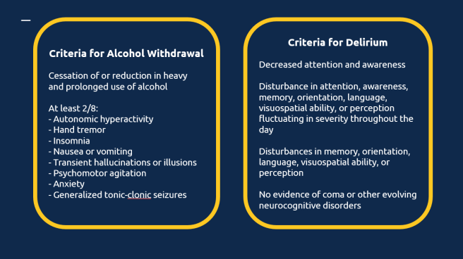
Figure 1. DSM-5 Criteria for Alcohol Withdrawal and Delirium. If the patient fulfills the criteria for both the diagnosis of alcohol withdrawal complicated by withdrawal delirium is made.

Pathophysiology

There are two different ethanol action in the central nervous system (CNS) that lead to symptoms of alcohol withdrawal. Overall, alcohol is a central nervous system depressant. It simultaneously increases inhibitory tone via modulation of GABA activity and decreases excitatory tone via modulation of excitatory amino acid activity. In a patient with alcohol abuse disorder, only a constant presence of alcohol keeps the necessary homeostasis. Sudden cessation unmasks the adaptive responses to chronic ethanol use, resulting in overactivity of the central nervous system.

Gamma-Aminobutyric acid (GABA) is the main inhibitory neurotransmitter in the brain. Highly specific binding sites for ethanol are found on the GABA receptor complex. Chronic ethanol use induces GABA receptor insensitivity to GABA resulting in a need for a stronger inhibitory stimulus to maintain a constant inhibitory tone. As alcohol tolerance develops, the individual retains arousal at alcohol concentrations that would normally produce lethargy or even coma in people who do not have alcohol use disorder. Sudden cessation of alcohol intake or a reduction from chronically elevated concentrations results in decreased inhibitory tone due to the lack of inhibitory effects of ethanol.

Glutamate is one of the major excitatory amino acids. When glutamate binds to the N-methyl-D-aspartate (NMDA) receptor, calcium influx leads to neuronal excitation by binding to the glycine receptor on the NMDA complex. Ethanol inhibits glutamate-induced excitation. Adaption occurs by increasing the number of glutamate receptors in an attempt to maintain a normal state of arousal.

Figure 2. Inhibitory and excitatory balance in a healthy individual.
Inhibitory and excitatory balance in an individual with chronic alcohol abuse.
Figure 3. Inhibitory and excitatory balance in an individual with chronic alcohol abuse. The constant presence of alcohol is needed to maintain inhibitory tone on insensitive GABA receptors and to inhibit excitatory tone on upregulated NMDA receptors.
Loss of the inhibitory and excitatory balance after alcohol cessation.
Figure 4. Loss of the inhibitory and excitatory balance after alcohol cessation. Upregulated NMDA receptors lead overexcitation, and insensitive GABA receptors are not enough to counteract that. Alcohol withdrawal symptoms ensue.

Differential Diagnosis

Alcohol withdrawal remains a clinical diagnosis. The severity of presentation can be assessed using a clinical assessment scale called Clinical Institute Withdrawal Assessment for Alcohol (CIWA-Ar) that can be found on MD Calc.

In some cases, several additional tests might be needed to rule out other conditions that mimic or coexist with alcohol withdrawal syndrome. This is especially true when the patient has altered mental status and fever. Conditions such as infection (e.g., meningitis), trauma (e.g., intracranial hemorrhage), metabolic abnormalities, drug overdose, hepatic failure, and gastrointestinal bleeding can mimic or coexist with alcohol withdrawal. Also, it is of marked importance to try to understand why the patient stopped consuming alcohol. If you establish that he wanted to get sober, that is great, you or the admitting team can help setting up rehab for him after the acute problems are controlled. However, you should get suspicious if there is not a clear cause for the abrupt cessation of alcohol intake since it could be an acute condition being masked by the withdrawal syndrome.

Initial workup might include:

  • Point of care glucose

  • CBC and platelets

  • Sodium, potassium, chloride, bicarbonate, BUN, creatinine

  • Calcium, magnesium, and phosphorus

  • Total protein, albumin, total bilirubin, AST, ALT, and alkaline phosphatase, and lipase

  • Creatine kinase

  • Chest x-ray to rule out simultaneous pneumonia

  • Consider head CT and lumbar puncture, if there are any findings concerning for trauma, intracranial hemorrhage, or CNS infections

  • Consider drug screen if concern for co-ingestion

Figure 5. The differential diagnosis for alcohol withdrawal. It is important to not anchor on this diagnosis and to look for mimics and conditions that might coexist with it.

Supportive Care

As important as proving control of the patient’s withdrawal symptoms is to provide high-quality supportive care, which includes:

  • Placement in a quiet and protective environment

  • Preference for chemical sedation over physical restraints, which should be removed as soon as adequate chemical sedation is achieved because resistance against restraints can increase temperature, produce rhabdomyolysis, and cause physical injury

  • IV fluids

  • Thiamine and glucose should be administered in order to prevent or treat Wernicke encephalopathy

  • Multivitamins containing or supplemented with folate should be given routinely

  • Deficiencies of glucose, potassium, magnesium, and phosphate should be corrected as needed

  • Nothing by mouth in the early stages of treatment to prevent aspiration

  • Patients considered at high risk for complications should be monitored in an intensive care unit

  • Consider ICU admission and EtCO2 monitoring in those patients with severe alcohol withdrawal per CIWA-Ar

Symptomatic Treatment

The basis for the treatment of alcohol withdrawal is CNS depressants, such as benzodiazepines, with a treatment goal of Richmond Agitation and Sedation Scale (RASS) -1 and HR < 110. No single drug benzodiazepine is superior to another. A common treatment strategy is to use a benzodiazepine of choice and give escalating doses until symptomatic control or until you reach criteria for refractory alcohol withdrawal.

Figure 6. Two common initial treatment strategies for alcohol withdrawal.

Refractory Withdrawal Delirium

Some patients have refractory delirium tremens (DT) despite treatment with high-dose benzodiazepines. Refractory DT is not clearly defined. It may be present if symptoms of severe withdrawal are not controlled adequately after the IV administration of more than 50 mg of diazepam or 10 mg of lorazepam during the first hour of treatment, or 200 mg of diazepam or 40 mg of lorazepam during the initial three to four hours of treatment. In such cases, as with any dangerous toxicologic disorder, you should obtain assistance from a medical toxicologist or poison control center. In case you diagnose your patient with alcohol withdrawal refractory to benzodiazepine treatment, you should have a few other options in your treatment arsenal.

Summary of sedation strategy.
Figure 7. Summary of sedation strategy. Initial treatment with benzodiazepines in escalating doses. If good response, keep regimen titrated to treatment goals. If no response, consider refractory withdrawal delirium and other pharmacologic options. If no response after secondary treatment, consider intubation with propofol.

Phenobarbital

There are case reports of up to 2000 mg of Phenobarbital administered orally or intravenously on the first day in patients with alcohol withdrawal delirium. Consider giving phenobarbital 130 to 260 mg IV, repeated every 15 to 20 minutes, until symptoms are controlled. Also, you can consider administering Phenobarbital earlier in the disease course. A randomized trial of 102 patients presenting to the emergency department with acute alcohol withdrawal, those treated with lorazepam and a single dose of Phenobarbital had substantially lower ICU admission rates compared with those treated with lorazepam alone (8 versus 25 percent).

Dexmetomedine

Another adjunctive medication for alcohol withdrawal is dexmedetomidine, an α2-adrenergic agonist that is used to provoke a state in which the patient is sedated but arousable, with a decreased sympathetic tone. Doses up to 0.7 μg per kilogram per hour have been administered in patients who do not have a good response to benzodiazepines. Heart block is a contraindication to this drug since it can cause bradycardia. In case it is given, blood pressure and heart rate must be closely monitored.

Propofol and Intubation

In patients who do not have a response to high doses of benzodiazepines (especially patients who are intubated), propofol may be administered to reach symptomatic control.

Take home points.
Figure 8. Take home points.

Further Reading

References

  • Isbell H, Fraser HF, Wilker A, et al. An experimental study of the etiology of rum fits and delirium tremens. Q J Stud Alcohol 1955; 16:1.
  • Mihic SJ, Ye Q, Wick MJ, et al. Sites of alcohol and volatile anaesthetic action on GABA(A) and glycine receptors. Nature 1997; 389:385.
  • Morrow AL, Suzdak PD, Karanian JW, Paul SM. Chronic ethanol administration alters gamma-aminobutyric acid, pentobarbital and ethanol-mediated 36Cl- uptake in cerebral cortical synaptoneurosomes. J Pharmacol Exp Ther 1988; 246:158.
  • Hoffman PL, Grant KA, Snell LD, et al. NMDA receptors: role in ethanol withdrawal seizures. Ann N Y Acad Sci 1992; 654:52.
  • Hecksel KA, Bostwick JM, Jaeger TM, Cha SS. Inappropriate use of symptom-triggered therapy for alcohol withdrawal in the general hospital. Mayo Clin Proc 2008; 83:274.
  • Hack JB, Hoffman RS. Thiamine before glucose to prevent Wernicke encephalopathy: examining the conventional wisdom. JAMA 1998; 279:583.
  • Hoffman RS, Goldfrank LR. The poisoned patient with altered consciousness. Controversies in the use of a ‘coma cocktail’. JAMA 1995; 274:562.
  • Hoffman RS, Goldfrank LR. Ethanol-associated metabolic disorders. Emerg Med Clin North Am 1989; 7:943.
  • Mainerova B, Prasko J, Latalova K, et al. Alcohol withdrawal delirium — diagnosis, course and treatment. Biomed Pap Med Fac Univ Palacky Olomouc Czech Repub 2013;157:1-9
  • Mayo-Smith MF, Beecher LH, Fischer TL, et al. Management of alcohol withdrawal delirium: an evidence-based practice guideline. Arch Intern Med 2004;164:1405-1412
  • Amato L, Minozzi S, Vecchi S, Davoli M. Benzodiazepines for alcohol withdrawal. Cochrane Database Syst Rev2010;3:CD005063-CD005063
  • Hjermø I, Anderson JE, Fink-Jensen A, Allerup P, Ulrichsen J. Phenobarbital versus diazepam for delirium tremens — a retrospective study. Dan Med Bull 2010;57:A4169-A4169
  • DeCarolis DD, Rice KL, Ho L, Willenbring ML, Cassaro S. Symptom-driven lorazepam protocol for treatment of severe alcohol withdrawal delirium in the intensive care unit. Pharmacotherapy 2007;27:510-518
  • DeBellis R, Smith BS, Choi S, Malloy M. Management of delirium tremens. J Intensive Care Med 2005;20:164-173
  • Cagetti E, Liang J, Spigelman I, Olsen RW. Withdrawal from chronic intermittent ethanol treatment changes subunit composition, reduces synaptic function, and decreases behavioral responses to positive allosteric modulators of GABAA receptors. Mol Pharmacol 2003; 63:53.
  • Nolop KB, Natow A. Unprecedented sedative requirements during delirium tremens. Crit Care Med 1985; 13:246.
  • Hack JB, Hoffmann RS, Nelson LS. Resistant alcohol withdrawal: does an unexpectedly large sedative requirement identify these patients early? J Med Toxicol 2006; 2:55.
  • Rosenson J, Clements C, Simon B, et al. Phenobarbital for acute alcohol withdrawal: a prospective randomized double-blind placebo-controlled study. J Emerg Med 2013; 44:592.
  • Rayner SG, Weinert CR, Peng H, Jepsen S, Broccard AF. Dexmedetomidine as adjunct treatment for severe alcohol withdrawal in the ICU. Ann Intensive Care 2012;2:12-12
  • Muzyk AJ, Fowler JA, Norwood DK, Chilipko A. Role of α2-agonists in the treatment of acute alcohol withdrawal. Ann Pharmacother 2011;45:649-657
  • Thomson AD, Cook CCH, Touquet R, Henry JA. The Royal College of Physicians report on alcohol: guidelines for managing Wernicke’s encephalopathy in the accident and emergency department. Alcohol Alcohol 2002;37:513-521
  • Koethe D, Juelicher A, Nolden BM, et al. Oxcarbazepine — efficacy and tolerability during treatment of alcohol withdrawal: a double-blind, randomized, placebo-controlled multicenter pilot study. Alcohol Clin Exp Res 2007;31:1188-1194
Cite this article as: Henrique Puls, Brasil, "A 32-year-old male with anxiety and tremor," in International Emergency Medicine Education Project, June 3, 2019, https://iem-student.org/2019/06/03/a-32-year-old-male-with-anxiety-and-tremor/, date accessed: April 27, 2024