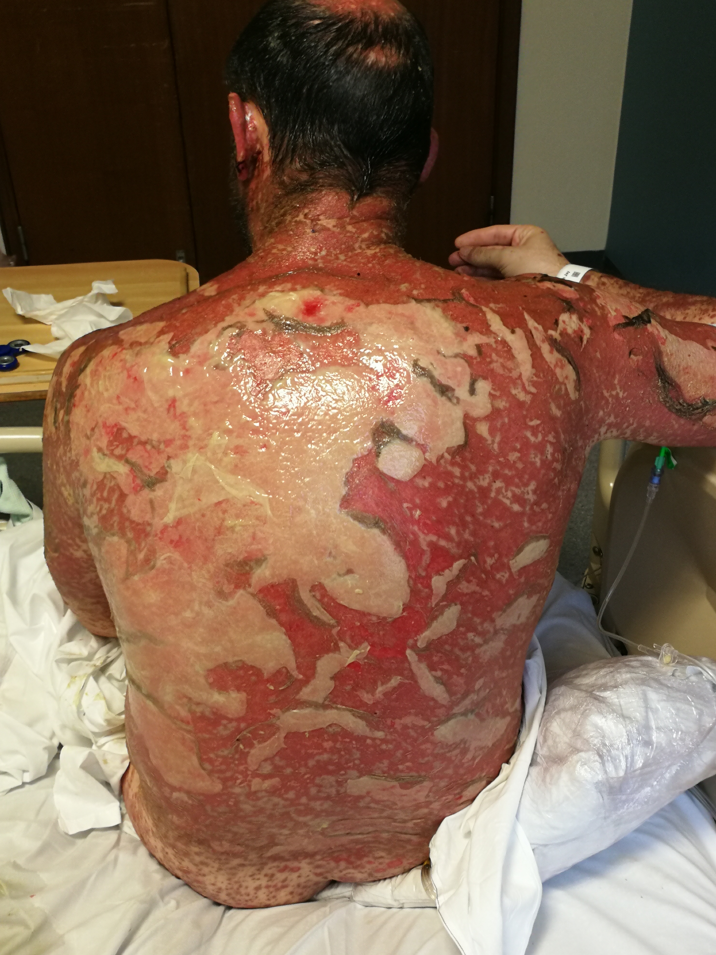
Dermatological emergencies : Stevens-Johnson Syndrome

International Emergency Medicine Education Project
We promote emergency medicine and provide free, reusable education resources for medical students and educators


Where This Attraction Come From? Emergency Medicine! It is maybe the most desired specialty all around the world. Countries are rapidly changing their systems to
Abdominal Aortic Aneurysm (AAA) Lit Sin Quek A 75-year-old obese man comes to the emergency department. He has history COPD, hypertension. He is a smoker
“Approach to poisoned patients” is one of the core EM clerkship topics! Poisonings Harajeshwar Kohli and Ziad Kazzi, USA An 18-year-old, previously healthy female, presents
Acute Mesenteric Ischemia Rabind Antony Charles, Singapore A 75-year-old woman presents to your Emergency Department (ED) with diffuse abdominal pain for the past day, associated
You may wonder “how to contribute” Promoting Emergency Medicine and improving undergraduate Emergency Medicine education (UEME) are the responsibility of all of us. We believe
This chapter describes how and why important the emergency medicine clerkship is. Although it aims to reach medical student/interns, there are many lessons to learn
Topic Today, we just wanted to emphasize a vital part of the suturing procedure which is sometimes forgotten. This is square knot. Simple, easy and important. Problem Suturing is
Gastrointestinal Bleeding by Moira Carrol, Gurpreet Mudan, and Suzanne Bentley, USA A 61-year-old man with a history of liver cirrhosis secondary to chronic EtOH abuse
Headache by Matevz Privsek and Gregor Prosen, Slovenia A 52-year old male comes to the ED with a severe headache. A triage nurse gives you
How ectopic pregnancy should be delivered to the students/interns. Clear, to the point! Ectopic Pregnancy by Dan O’Brien, USA A 24-year-old woman presents to the
In case you did not encounter a flail chest today. https://youtu.be/k78yENIpmFE
Acute Appendicitis by Ozlem Dikme, Turkey A previously healthy 22-year-old male was brought to the emergency department (ED) with recently-started abdominal pain. He had not
In case you didn’t encounter shortness of breath today! Go To “Respiratory Distress” Chapter Go To “Chest Pain” Chapter iEM Education Project Team uploads many
In case you didn’t encounter abdominal pain today! Go To “Abdominal Pain” Chapter iEM Education Project Team uploads many clinical picture and videos to the


