Approach to Acutely Confused Patient (2025)

by Mehnaz Zafar Ali

You Have New Patients!

Patient 1

You meet a 40-year-old man in the ED, held by three security staff, looking diaphoretic and agitated, having tachycardia, and pointing vaguely in a direction as if interacting with imaginary people. When you try to assess him, he appears to be confused and disoriented and smells of alcohol. Over 6 hours, the patient has tremulousness, gets easily frightened, and gets further uncooperative for examination.

The image was produced by using ideogram 2.0.

Patient 2

You evaluate an 80-year-old woman in the ICU. She has a history of diabetes mellitus, hypertension, depression, and a stroke two years ago. She was admitted due to increased sleepiness, urinary and fecal incontinence for one week, and difficulty recognizing people. Before her admission, she was active and independent, had a reasonably good memory, and could manage household responsibilities. On physical examination, her eyes remain spontaneously closed but open with audible stimuli, and she is disoriented to time, place, and person.

The image was produced by using ideogram 2.0.

Introduction

Delirium is a rapidly developing clinical syndrome characterized by alterations in attention, consciousness, and awareness, with a reduced ability to focus, sustain, or shift attention. It commonly occurs in the elderly, with an incidence reported in 10% to 30% of patients hospitalized for medical illnesses and up to 50% following high-risk procedures [1].

This condition is also referred to as acute organic brain syndrome, characterized by rapid onset, diurnal fluctuations, and a duration of less than six months. Its behavioral presentation can vary, with the following manifestations.

  • Hyperactive Delirium: Patients present with increased agitation and heightened sympathetic activity. They may exhibit hallucinations, delusions, and combative or uncooperative behavior.

  • Hypoactive Delirium: Patients display increased somnolence and reduced arousal. The diagnosis is often overlooked due to its subtle clinical manifestations, which are frequently mistaken for fatigue or depression. This subtype is associated with higher rates of morbidity and mortality.

  • Mixed Presentation: Patients fluctuate between hyperactive and hypoactive delirium.

Delirium tremens (DT) is the most severe form of alcohol withdrawal syndrome and can be fatal. It typically occurs within 2 to 4 days following complete or significant abstinence from heavy alcohol consumption in approximately 5% of patients, with mortality rates as high as 50%. Alcohol functions as a depressant, similar to benzodiazepines and barbiturates, and affects serotonin and gamma-aminobutyric acid type A (GABA A) receptors, leading to tolerance and habituation.

Delirium is a dangerous and often preventable condition, associated with significant costs and increased morbidity and mortality. Among delirium patients presenting to the emergency department, there is a 70% increased risk of death within six months. In the ICU, delirium is linked to a 2- to 4-fold increased risk of overall mortality. Prevention, early diagnosis, and treatment of the underlying cause, along with well-coordinated care, are essential to improve patient outcomes.

General Approach

The diagnosis of delirium is primarily clinical and relies on careful history-taking, mental status examination, and detailed cognitive assessment. While laboratory and diagnostic tests may assist in identifying the underlying etiology, the initial evaluation should focus on addressing reversible causes. Life-threatening conditions must be promptly recognized, requiring rapid intervention and stabilization.

Differential Diagnoses

Delirium can present with symptoms that may be easily mistaken for mental illness, such as acute aggression, irritability, restlessness, and visual hallucinations [1]. Delirium mimics may include psychosis or mood disorders in the case of hyperactive delirium, and depression in the case of hypoactive delirium.

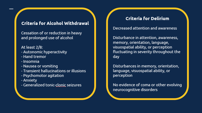
According to the International Classification of Diseases (ICD-10) guidelines [2], a definite diagnosis of delirium requires the presence of symptoms (mild or severe) in each of the five described areas. These include: impairment of consciousness and attention (ranging from clouding to coma, with a reduced ability to direct, focus, sustain, and shift attention), global disturbance of cognition, psychomotor disturbances, disturbance of the sleep-wake cycle, and emotional disturbances.

Delirium

Delirium typically presents with an acute onset and progresses rapidly. It often resolves completely with treatment of the underlying cause. Clinically, it is characterized by fluctuating levels of consciousness, inattention, disorientation, worsening symptoms in the evening (a phenomenon known as sundowning), and transient visual hallucinations. Delirium carries significant risks, including high mortality due to the underlying medical condition, as well as increased risk of falls, injuries, exhaustion, or aggression.

Dementia

Dementia has an insidious onset and follows a chronic, progressive course marked by continuous deterioration over time. Key clinical features include memory disturbances, changes in personality or behavior, apathy, and apraxia. Individuals with dementia are at risk of falls, neglect, abuse, agitation, and wandering away from their safe environments.

Depression

Depression typically has a slow onset and an episodic course, with periods of remission and recurrence. Symptoms include a persistently depressed mood, loss of interest or pleasure in activities, reduced energy, feelings of hopelessness, disturbances in sleep and appetite, difficulties with concentration, and pervasive negative thoughts, often accompanied by guilt. The associated risks include suicide, deliberate self-harm, neglect, and agitation.

Psychosis

Psychosis usually begins insidiously and follows a progressive course punctuated by episodes of exacerbation. Clinical features include delusions, auditory hallucinations, disorganized thoughts, social withdrawal, apathy, avolition (lack of motivation), and impaired reality testing. Psychosis poses risks such as aggression, harm to others, and non-adherence to treatment, which can exacerbate the condition further.

History and Physical Examination Hints

It is of paramount importance to obtain a detailed corroborative history regarding the onset, course, and progression of the illness, along with performing a thorough physical and neurological examination of the patient. A biopsychosocial formulation must identify the predisposing, precipitating, and perpetuating causes of delirium [1].

The mnemonic “I WATCH DEATH,” developed by Dr. M.G. Wise in 1986, is a valuable tool for clinicians to screen for possible causes of delirium [3].

  • I – Infections: Infections are a common cause and can include conditions such as sepsis, urinary tract infections, encephalitis, and meningitis.

  • W – Withdrawal: Sudden withdrawal from substances such as alcohol, sedatives, or drugs can lead to significant medical complications.

  • A – Acute Metabolic Disturbances: Issues such as electrolyte imbalances (e.g., hyponatremia) and organ failure, such as hepatic or renal failure, can significantly disrupt normal physiological functions.

  • T – Trauma: Physical injuries, including head trauma and falls, are notable causes that may lead to further complications like bleeding or swelling.

  • C – CNS Pathology: Central nervous system disorders such as stroke, hemorrhage, seizures, or the presence of space-occupying lesions like tumors can have profound impacts on a patient’s condition.

  • H – Hypoxia: A lack of adequate oxygen supply, often due to anemia or hypotension, can result in significant systemic effects.

  • D – Deficiencies: Nutritional deficiencies, particularly a lack of essential vitamins and minerals like thiamine, can result in various clinical symptoms.

  • E – Endocrine Disorders: Hormonal imbalances, including thyroid storm and hyperglycemia, can disrupt metabolic processes and cause severe systemic effects.

  • A – Acute Vascular Events: Sudden vascular events, such as subarachnoid hemorrhage, require prompt identification and management due to their life-threatening nature.

  • T – Toxins or Drugs: Exposure to industrial poisons, carbon monoxide, or drugs with anticholinergic properties can have toxic effects on the body.

  • H – Heavy Metal Poisoning: Exposure to heavy metals such as lead and mercury can lead to chronic toxicity and require specific interventions.

Several factors increase the likelihood of developing delirium, especially in vulnerable populations:

  1. Age: Both elderly individuals and young children are at heightened risk due to their increased susceptibility to physiological and cognitive changes.

  2. Recent Hospitalizations: Hospital stays, particularly those involving medical illnesses or surgical procedures, can act as significant stressors and predispose individuals to delirium.

  3. Pre-existing Brain Conditions: Conditions like brain damage or dementia further increase the risk, as they impair cognitive resilience.

  4. Chronic Medical Disorders: Long-term health conditions often contribute to a state of chronic physiological stress, increasing the likelihood of delirium.

  5. Sensory Deprivation: Impairments in vision or hearing can lead to sensory deprivation, which may exacerbate confusion and disorientation.

  6. Substance Use Disorders: Alcohol or drug use disorders are major contributors to the onset of delirium, particularly during withdrawal periods or intoxication.

  7. Medications: The use of psychotropic medicines and polypharmacy (simultaneous use of multiple medications) heightens the risk of delirium due to potential drug interactions and side effects.

  8. History of Delirium: Individuals with a previous history of delirium are more likely to experience recurrent episodes, particularly if the underlying risk factors persist.

  9. Malnutrition: Poor nutritional status can exacerbate vulnerability to delirium by impairing metabolic and neurological functions.

  10. Burns: Severe burns create systemic inflammation and stress, which can predispose individuals to delirium.

Screening tools for delirium, such as the Mini-Mental Status Examination (MMSE) [4] and the Confusion Assessment Method (CAM) [5], are valuable for early identification and intervention. These tools can also be used to monitor clinical improvement when performed repeatedly during the course of the illness.

The Confusion Assessment Method (CAM) includes four key features to identify delirium. A diagnosis of delirium requires the presence of Features 1 and 2 and either Feature 3 or Feature 4:

Feature 1 – Acute Onset and Fluctuating Course: There is evidence of an acute change in mental status from the patient’s baseline.
The abnormal behavior fluctuates throughout the day, tending to come and go or change in severity.

Feature 2 – Inattention: The patient has difficulty focusing attention, is easily distractible, or cannot keep track of what is being said.

Feature 3 – Disorganized Thinking: The patient demonstrates disorganized or incoherent thinking, such as rambling or irrelevant conversation, illogical flow of ideas, or unpredictable switching between subjects.

Feature 4 – Altered Level of Consciousness: The patient’s consciousness level deviates from “alert.” It may range from hyperalert (vigilant) to lethargy, stupor, or coma.

The CAM is a widely used, reliable tool with high sensitivity (94–100%) and specificity (90–95%). It enables quick and accurate identification of delirium, facilitating early intervention to manage underlying causes and improve patient outcomes.

Confusion Assessment Method (CAM) Instrument:

  1. Acute Onset:
    • This involves an abrupt change in the patient’s mental status, which is evident when comparing their current state to their baseline cognitive function. This change may be noticed by family members, caregivers, or clinicians and is typically indicative of an acute underlying medical issue or condition.
  2. Inattention:
    • 2A: The patient has difficulty concentrating or paying attention. This may manifest as being easily distracted, unable to follow conversations, or losing track of what is being discussed.
    • 2B: If inattention is present, the behavior often fluctuates over time, meaning it can improve or worsen during an assessment or throughout the day.
  3. Disorganized Thinking:
    • The patient’s thought process appears chaotic or incoherent. They may exhibit rambling, irrelevant speech, an illogical sequence of ideas, or rapid, unpredictable topic changes during a conversation. This suggests a loss of organized, goal-directed thinking.
  4. Altered Level of Consciousness:
    • The patient’s alertness deviates from normal. This can range from:
      • Alert (normal): Fully awake and responsive.
      • Vigilant (hyperalert): Overly sensitive to stimuli, easily startled, or hypervigilant.
      • Lethargic: Drowsy but easily aroused.
      • Stupor: Difficult to arouse, with limited responsiveness to stimuli.
      • Coma: Unarousable and non-responsive.
  5. Disorientation:
    • The patient is confused about time, place, or identity. They may incorrectly believe they are in a different location, misjudge the time of day, or demonstrate an inability to recognize familiar surroundings or people.
  6. Memory Impairment:
    • Memory issues are evident when the patient cannot recall recent events, forgets instructions, or struggles to remember details of their hospital stay or interactions.
  7. Perceptual Disturbances:
    • The patient may experience hallucinations (e.g., seeing or hearing things that aren’t present), illusions (misinterpreting real stimuli, such as mistaking a shadow for an object), or misinterpretations (believing something benign, such as a coat rack, is threatening).
  8. Psychomotor Disturbances:
    • 8A (Agitation): The patient may exhibit increased motor activity, such as restlessness, repeatedly picking at bedclothes, tapping their fingers, or making frequent, sudden movements.
    • 8B (Retardation): Alternatively, the patient may show decreased motor activity, appearing sluggish, staring into space, staying in the same position for extended periods, or moving very slowly.
  9. Altered Sleep-Wake Cycle:
    • Disturbances in the patient’s sleep pattern are evident. They may experience excessive daytime sleepiness coupled with difficulty sleeping at night, or their sleep-wake rhythm may become reversed.

Associated Features

Certain medical conditions can present with a range of distressing symptoms and features:

  1. Hallucinations and Illusions: Patients may experience vivid and often frightening visual or auditory hallucinations. Additionally, tactile hallucinations, such as the sensation of insects crawling on the body, can occur, adding to their distress.

  2. Autonomic Disturbances: Marked autonomic instability is common and may include symptoms such as tachycardia, fever, hypertension, sweating, and pupillary dilation.

  3. Psychomotor and Coordination Issues: Psychomotor agitation and ataxia (lack of muscle coordination) are frequently observed, contributing to physical instability and difficulty performing tasks.

  4. Sleep Disturbances: Insomnia is a notable feature, often accompanied by a reversal of the sleep-wake cycle, further exacerbating cognitive and physical impairments.

It is crucial to obtain a detailed history of the patient’s premorbid personality, as this helps establish their baseline cognitive state and allows the clinician to determine the magnitude of cognitive deterioration. Patients with fluctuating levels of consciousness may experience rapid shifts in their activity levels, ranging from extreme psychomotor excitement to sleepiness during an interview [1].

The Mental State Examination (MSE) should include an assessment of mood (e.g., apathy, blunted affect, emotional lability), behavior (e.g., withdrawn, agitated), activity levels, thoughts (e.g., delusions), and perceptions (e.g., hallucinations, illusions). A brief cognitive assessment may utilize the COMA framework, which evaluates Concentration, Orientation, Memory, and Attention.

Clinical Institute Withdrawal Assessment of Alcohol Scale, Revised (CIWA-Ar)

The CIWA-R is a tool designed to standardize the assessment of withdrawal severity in patients experiencing alcohol withdrawal. This instrument is particularly useful for guiding treatment decisions and ensuring appropriate management of symptoms.

Alcohol withdrawal delirium progresses through distinct stages, including:

  • Tremulousness or Jitteriness: Occurs within 6–8 hours of cessation or reduction in alcohol use.
  • Psychosis and Perceptual Symptoms: Develops between 8–12 hours, marked by hallucinations and disorganized thinking.
  • Seizures: Typically occur within 12–24 hours of withdrawal.
  • Delirium Tremens: The most severe stage, manifesting within 24–72 hours and potentially lasting up to one week. This phase is characterized by confusion, autonomic instability, and significant risk of complications.

The CIWA-R plays a critical role in monitoring these stages and ensuring timely interventions to mitigate risks associated with alcohol withdrawal.

Click here to download full CIWA-R evaluation form.

Diagnostic Tests and Interpretation

Relevant laboratory tests and diagnostic imaging are recommended to assess the underlying etiology of delirium. Routine workups for electrolytes, kidney and liver function, and pregnancy tests for women are advised. Blood tests can help identify medical conditions that may mimic delirium, such as hypoglycemia and diabetic ketoacidosis (via blood sugar levels) or thyrotoxicosis (via thyroid profile). Test results indicative of long-term heavy alcohol use, such as evidence of cirrhosis or liver failure on ultrasound, macrocytic anemia, and elevated liver transaminase levels—particularly gamma-glutamyl transpeptidase—can aid in reaching the correct diagnosis [6].

Positron emission tomographic (PET) studies have suggested a globally low rate of metabolic activity, particularly in the left parietal and right frontal areas, in otherwise healthy individuals withdrawing from alcohol. Diffuse slowing of the background rhythm has been observed on electroencephalography (EEG) in patients suffering from acute delirium, except in cases of alcohol-related delirium tremens, which typically exhibit fast activity [1].

Management

Delirium is a medical emergency requiring immediate hospitalization to correct the underlying causes while minimizing risks associated with behavioral symptoms, aggression, dehydration, falls, and injury. High-potency antipsychotics in low doses are recommended for managing aggression and behavioral symptoms. Haloperidol (Haldol) has been extensively studied for reducing agitation due to delirium [7]. Evidence also supports the use of other atypical antipsychotics such as risperidone. Aripiprazole has demonstrated significant benefit in the complete resolution of hypoactive delirium [8].

The use of benzodiazepines should be restricted to cases of delirium caused by alcohol withdrawal. If liver function is not impaired, a long-acting benzodiazepine, such as chlordiazepoxide or diazepam, is preferred and can be administered orally or intravenously. In cases of reduced liver function, lorazepam may be given orally or parenterally as needed to stabilize vital signs and sedate the patient. These medications should then be tapered gradually over several days with close monitoring of vital signs. Anticonvulsants like carbamazepine and valproic acid are also effective in managing alcohol withdrawal. However, antipsychotics should be avoided in such cases due to their potential to lower the seizure threshold. Chronic alcoholics are at high risk of vitamin B1 (thiamine) deficiency, which can predispose them to Wernicke-Korsakoff syndrome (characterized by memory problems, confabulation, and apathy), cerebellar degeneration, and cardiovascular dysfunction. To mitigate this risk, such patients should receive 100 mg of thiamine intravenously before glucose administration.

Environmental modification strategies are particularly useful for managing delirious patients. These include providing well-illuminated rooms with good ventilation and reorientation cues such as calendars and alarm clocks. Assigning patients to a room near the nursing station allows for closer monitoring, ideally with the presence of a family member or close friend. In severe cases with agitation or injury risk, one-on-one supervision is advisable to ensure patient safety [1]. Both under-stimulation and overstimulation should be avoided. The use of physical restraints should be considered a last resort, with frequent monitoring and discontinuation as soon as possible. Psychoeducation for family members and caregivers is crucial to manage expectations and improve their involvement in the patient’s care [2].

Special Patient Groups and Other Considerations

Elderly patients are at high risk of altered mental status, and studies have recommended advanced age as an independent risk factor warranting screening of this vulnerable group through structured mental state assessments. It is important to recognize that behavioral manifestations of this magnitude should not be regarded as a normal part of the aging process. Dementia must be carefully differentiated from delirium in the geriatric population, as dementia typically presents with an insidious onset and a progressive course [3].

Other risk factors in the elderly that require attention include underlying neurological causes, multiple medical comorbidities, polypharmacy, poor drug metabolism, and sensory limitations [9]. Medications for elderly patients should be initiated at lower doses, and potential drug interactions must be considered whenever new medications are introduced.

The pediatric age group may present with nonspecific symptoms of acute onset, necessitating a detailed history and physical examination to rule out causes such as fever, injury, or foreign objects. Pregnancy, meanwhile, may predispose healthy women to medical conditions such as diabetes, venous thromboembolism, strokes, and eclampsia [9].

When To Admit This Patient

Admission decisions for confused patients or those undergoing alcohol withdrawal require a multifaceted approach that prioritizes accurate diagnosis, evidence-based treatment, and legal considerations. These decisions should aim to address the immediate medical needs while planning for long-term recovery and safety.

Admitting a confused patient requires careful evaluation of the underlying causes, as confusion can result from various conditions such as dementia, delirium, or depression, each requiring distinct management strategies [10]. Delirium, an acute confusional state, is particularly prevalent in older adults and often develops rapidly with fluctuating severity [11]. It is essential to determine whether the confusion is acute, chronic, or a combination of both, as this distinction guides the initial management plan [11].

Risk factors for acute confusion include admission from non-home settings, lower cognitive scores, restricted activity levels, infections, and abnormal laboratory values. These indicators suggest frailty and may also point to underlying chronic undernutrition or dehydration [12]. Early recognition and appropriate management are crucial to reducing morbidity and mortality, as confusion is often misdiagnosed or undertreated in hospital settings [10].

Furthermore, legal and ethical challenges, such as evaluating a patient’s decision-making capacity and ensuring that any necessary restraints are lawful and ethical, must be addressed to avoid infringing on the patient’s rights [13]. A comprehensive assessment of cognitive and physical status, coupled with an understanding of legal considerations, is essential for developing a management plan that effectively addresses the specific causes and risks associated with confusion [11-13].

Disposition decisions for confused patients, including those undergoing alcohol withdrawal, require a comprehensive and systematic approach that integrates accurate diagnosis, appropriate treatment, and continuous monitoring. Alcohol withdrawal can result in severe complications, such as seizures and delirium tremens, with mortality rates ranging from 1% to 30%, depending on the quality of treatment provided [14]. Prompt identification and management are critical, often involving benzodiazepines like diazepam to alleviate symptoms and prevent progression to life-threatening conditions [15]. Management becomes particularly challenging in critically ill patients, as incomplete alcohol consumption histories and the need for adjunctive medications beyond benzodiazepines complicate care during severe withdrawal or delirium tremens [16].

Emergency departments frequently encounter substance use disorders; however, less than half of alcohol-related issues are identified, highlighting the importance of comprehensive assessments and evidence-based interventions. Effective disposition decisions rely on early identification, tailored treatment strategies, and ongoing evaluations to ensure patient safety and recovery.

Clinical Pearls

  • Alcohol Withdrawal Characteristics: Alcohol withdrawal can begin within hours to days following heavy and prolonged alcohol use. A key feature of alcohol withdrawal is autonomic hyperactivity, which may present as increased heart rate, sweating, tremors, and other signs of sympathetic nervous system overactivity.
  • Overlap with Sedative-Hypnotic Withdrawal: The diagnostic criteria and symptoms for alcohol withdrawal are identical to those for sedative-hypnotic withdrawal. This similarity highlights the importance of carefully assessing a patient’s history of substance use to guide appropriate management.
  • Treatment Approaches:
    • Delirium Due to General Medical Conditions: The preferred treatment is low doses of high-potency antipsychotics, which help manage symptoms without excessive sedation or complications.
    • Alcohol Withdrawal: Benzodiazepines remain the first-line treatment to alleviate withdrawal symptoms and prevent complications such as seizures or delirium tremens. In cases where hepatotoxicity is a concern, short-acting benzodiazepines like lorazepam are preferred due to their safer profile in patients with compromised liver function.
  • Hallucinations and Diagnosis: Visual hallucinations are more characteristic of delirium than of primary psychiatric disorders. This distinction is critical in differentiating between medical and psychiatric causes of altered mental status.

Revisiting Your Patient

Patient 1

The image was produced by using ideogram 2.0.

The patient presents with the smell of alcohol and clinical features consistent with delirium tremens, a severe manifestation of alcohol withdrawal.

Further Management: The patient should be treated promptly with a benzodiazepine, starting with high doses and tapering as recovery progresses. Chronic alcohol users are commonly deficient in vitamin B1 (thiamine), which can result in dementia and cognitive impairments. Thiamine replacement should be administered prior to glucose to prevent the development of Wernicke-Korsakoff syndrome [17].

Patient 2

The image was produced by using ideogram 2.0.

The patient is unresponsive to stimuli, disoriented, and has multiple medical conditions, which is suggestive of delirium due to a general medical condition, hypoactive type.

Further Management: Immediate steps should include ensuring 24-hour supervision, investigating the underlying cause, and implementing reorientation strategies. Low-dose antipsychotics have been recommended, with studies reporting complete resolution of symptoms with the use of aripiprazole and other atypical antipsychotics [18].

Author

Picture of Mehnaz Zafar Ali

Mehnaz Zafar Ali

Consultant Psychiatrist, Al Amal Psychiatry Hospital, Emirates Health Services, Dubai, United Arab Emirates

Listen to the chapter

References

  1. Gleason OC. Delirium. Am Fam Physician. 2003;67(5):1027-1034.
  2. World Health Organization. Organic, including symptomatic, mental disorders. In: International Statistical Classification of Diseases and Related Health Problems. 10th ed. 2016:182-188.
  3. Gower LE, Gatewood MO, Kang CS. Emergency department management of delirium in the elderly. West J Emerg Med. 2012;13(2):194-201. doi:10.5811/westjem.2011.10.6654.
  4. Folstein MF, Folstein SE, McHugh PR.Mini-mental state”. A practical method for grading the cognitive state of patients for the clinician. J Psychiatr Res. 1975;12(3):189-198. doi:10.1016/0022-3956(75)90026-6.
  5. Inouye SK, van Dyck CH, Alessi CA, Balkin S, Siegal AP, Horwitz RI. Clarifying confusion: the confusion assessment method. A new method for detection of delirium. Ann Intern Med. 1990;113(12):941-948. doi:10.7326/0003-4819-113-12-941.
  6. Chan M, Moukaddam N, Tucci V. Stabilization and management of the acutely agitated or psychotic patient. In: Cevik AA, Quek LS, Noureldin A, Cakal ED, eds. International Emergency Medicine Education Project. 1st ed. iEM Education Project; 2018:452-457.
  7. Smit L, Slooter AJ, Devlin JW, et al. Efficacy of haloperidol to decrease the burden of delirium in adult critically ill patients: the EuRIDICE randomized clinical trial. Crit Care. 2023;27(1):413. doi:10.1186/s13054-023-04692-3.
  8. Lodewijckx E, Debain A, Lieten S, et al. Pharmacologic treatment for hypoactive delirium in adult patients: a brief report of the literature. J Am Med Dir Assoc. 2021;22(6):1313-1316.e2. doi:10.1016/j.jamda.2020.12.037.
  9. Cetin M, Oktem B, Canakci ME. Altered mental status. In: Cevik AA, Quek LS, Noureldin A, Cakal ED, eds. International Emergency Medicine Education Project. 1st ed. iEM Education Project; 2018:111-121.
  10. Winstanley L, Glew S, Harwood RH. A foundation doctor’s guide to clerking the confused older patient. Br J Hosp Med (Lond). 2010;71(5):M78-M81. doi:10.12968/hmed.2010.71.Sup5.47934.
  11. Andrews H, Clarke A, Parmar S, et al. You’ve been bleeped: the confused patient. BMJ. 2015;351:h3266. doi:10.1136/sbmj.h3266.
  12. Wakefield BJ. Risk for acute confusion on hospital admission. Clin Nurs Res. 2002;11(2):153-172. doi:10.1177/105477380201100205.
  13. Lyons D. The confused patient in the acute hospital: legal and ethical challenges for clinicians in Scotland. J R Coll Physicians Edinb. 2013;43(1):61-67. doi:10.4997/jrcpe.2013.114.
  14. Thanyanuwat R. Patients who suffer from alcohol withdrawal and disorientation. J Med Assoc Thai. 2013;96(2):78-83.
  15. Thompson WL. Management of alcohol withdrawal syndromes. Arch Intern Med. 1978;138(2):278-283. doi:10.1001/archinte.1978.03630260068019.
  16. Sutton LJ, Jutel A. Alcohol withdrawal syndrome in critically ill patients: identification, assessment, and management. Crit Care Nurse. 2016;36(1):28-40. doi:10.4037/ccn2016420.
  17. Toy EC, Klamen DL. Alcohol withdrawal. In: Case Files: Psychiatry. 6th ed. McGraw-Hill Education; 2020:400-405.
  18. Lodewijckx E, Debain A, Lieten S, et al. Pharmacologic treatment for hypoactive delirium in adult patients: a brief report of the literature. J Am Med Dir Assoc. 2021;22(6):1313-1316.e2. doi:10.1016/j.jamda.2020.12.037.

Reviewed and Edited By

Picture of Arif Alper Cevik, MD, FEMAT, FIFEM

Arif Alper Cevik, MD, FEMAT, FIFEM

Prof Cevik is an Emergency Medicine academician at United Arab Emirates University, interested in international emergency medicine, emergency medicine education, medical education, point of care ultrasound and trauma. He is the founder and director of the International Emergency Medicine Education Project – iem-student.org, chair of the International Federation for Emergency Medicine (IFEM) core curriculum and education committee and board member of the Asian Society for Emergency Medicine and Emirati Board of Emergency Medicine.

Delirium and Dementia (2024)

by Lo Lucian Simeon, Ngai Oona Wing Yan, & Lo Yat Hei

You have a new patient!

Adam is a 76-year-old man who is brought to the emergency room by his family members, complaining of a lack of responsiveness and general lethargy. According to his family, Adam has been having increasing memory problems in the past year and has gotten lost while walking around his neighborhood multiple times. His personality has changed and becomes agitated easily. He is also becoming less attentive to personal hygiene, wearing dirty clothes for several days, and having several episodes of urinary incontinence. Today, his family members noted that he had fallen asleep multiple times and showed no interest in his food. He did not respond when addressed by name. At the time of presentation, he is conscious, but appears lethargic and uncooperative. He cannot tell where he is and does not seem to recognize his family members. His past medical history includes hypertension and hypercholesterolemia. He is taking amlodipine and simvastatin.

Vitals show a heart rate of 108 beats per minute, blood pressure 154/84 mmHg, temperature 36.7℃, respiratory rate 20 breaths per minute, and an oxygen saturation of 98% on room air. His Glasgow coma score is E4V4M6.

What do you need to know?

Importance

Dementia and delirium are two medical conditions that significantly impact the health and well-being of older adults and their families. In this case, Adam’s symptoms suggest that he may be experiencing one or both of these conditions, and it is important to understand their relevance in clinical practice.

Dementia is defined as an acquired global decline in cognitive function, affecting one’s memory, language, learning, and behavior without impairment of consciousness. Dementia is associated with a gradual, progressive decline. It is a leading cause of disability and dependence among older adults, with advancing age being one of the most significant risk factors [1]. With the global population aging, the number of individuals living with dementia is expected to rise significantly. The World Health Organisation estimates that 47 million people worldwide live with dementia, and this number is expected to triple by 2025 [2].

Delirium, conversely, is a clinical syndrome characterized by an acute state of confusion, inattention, and cognitive impairment. It can occur in people of any age, but is particularly common among the older population and hospitalized patients [1]. Delirium can wax and wane over time, unlike dementia, which is more progressive and persistent.

Dementia and delirium pose a tremendous burden not only on patients and caregivers, but also on our healthcare system and society. Therefore, understanding the significance of dementia and delirium is crucial in clinical practice. Identifying and managing these conditions early improves clinical outcomes and optimizes quality of life.

Epidemiology & Pathophysiology

Dementia is a condition that is more commonly seen in older individuals, with the incidence increasing from the age of 65.  An exception is frontotemporal dementia, a rare type of dementia that is usually diagnosed from the age of 40 to 60. The most prevalent type of dementia is Alzheimer’s disease, which accounts for 60-80% of all cases. Other neurodegenerative dementias, such as vascular dementia, dementia due to Lewy bodies, Parkinson’s disease, and frontotemporal dementia, account for the remaining cases [3].

The underlying pathophysiology of dementia varies depending on the type and subtype, with most types involving damage to neurons and their connections in the brain. Abnormal protein accumulation is a common feature for many types of dementia, including amyloid and tau in Alzheimer’s disease, Lewy bodies with alpha-synuclein protein in Lewy body disease, and mutations causing the deposition of TDP-43 and tau proteins in frontotemporal dementia. Other factors, such as ischemic injury, HIV infection, and alcohol consumption, can also lead to cytotoxic processes in the brain and contribute to the development of dementia [4].

On the other hand, the epidemiology of delirium is more complex as it varies depending on age and underlying medical conditions. Although delirium is more prevalent in older individuals, with rates increasing after the age of 70, it is also common in younger patients suffering from chronic illnesses such as cardiovascular and renal comorbidities, dementia, or psychiatric illnesses [5].

The pathophysiology of delirium can result from various physiological and structural lesions in the brain. While its mechanisms are not fully understood, delirium can be caused by neurotransmitter imbalances, brain lesions involving the ascending reticular activating system, as well as disrupted blood-brain barrier function that causes the leakage of neurotoxic agents into the brain. Patients with impaired cholinergic transmission, such as those with Alzheimer’s disease, are particularly susceptible to delirium caused by medication use. Additionally, delirium can result from alcohol abuse, drug withdrawal, mental illnesses, psychosocial stress, and sleep deprivation [6].

Epidemiological and pathophysiological data on dementia and delirium allow physicians to identify individuals at risk and intervene appropriately. Since the development of dementia and delirium are multifactorial and the pathophysiology is variable among patients, evaluating and treating delirium and dementia is based on clinical gestalt and the presumed underlying cause.

Medical History

Dementia and delirium are two diagnoses that must be considered in elderly patients presenting with cognitive change to the emergency department. Differentiation between the two conditions is based on features noted in the history and physical examination. Table 1 lists symptoms that can help differentiate between the two conditions.

Delirium typically presents with sudden onset of impaired awareness, confusion, clouding of consciousness, and disturbances of perception (e.g., illusions or hallucinations). Delirium should be suspected when there is an acute deterioration in behavior, cognition, and daily functioning [7]. Delirious patients usually have short-term memory issues and may be disoriented by time and place. Abnormalities of cognition and behavior can fluctuate over brief periods. The level of awareness may range from hypervigilant and agitated to blunt and unreactive. The patient’s speech may be incoherent, nonsensical, or tense. The patient usually has no discernible focal neurological defect [8].

Dementia has various presentations according to the specific types, but symptoms often overlap. Alzheimer’s dementia, the most common type, presents with a history of a chronic, steady decline in cognitive ability, especially memory. It is often associated with difficulties in social relationships, activities of daily living, and work. During the early stages of dementia, clinical presentations can be quite subtle, and patients may try to hide their cognitive impairments [9].

Patients who present to the emergency department with symptoms of dementia are most likely in the later stages of disease progression. Acute presentation of dementia is possible in vascular dementia, and this subtype may present with symptoms of focal neurological deficit. Patients who are demented typically do not present with any impairment of consciousness. However, acute episodes of delirium can be superimposed on patients who have dementia.  For example, Lewy body dementia can present with fluctuating levels of consciousness. Diagnosing uncommon variants poses a challenge to emergency physicians and is often done only after referral to a neurologist [10].

Obtaining a thorough history is essential in diagnosing delirium and dementia. Unfortunately, delirious or demented patients are often disoriented and cognitively impaired, resulting in the inability to provide accurate information about their condition. A detailed history should be obtained from family, caregivers, and healthcare staff (nurses, healthcare assistants, and other allied health professionals).

Important features to note during history are the onset of symptoms, factors that worsen or improve symptoms, drug or alcohol use, pre-existing endocrine or psychiatric disorders, exposure to toxins or traumatic injury, social history, and previous similar episodes of confusion or altered mental status. Drug history is particularly important as the use of drugs that impair cognition (e.g., analgesics, anticholinergics, psychotropic medications, and sedatives) may explain the presenting symptoms. 

Determining the onset of symptoms, in particular, for patients with dementia, can be difficult due to the gradual nature of the disease. Questions like, “When did you first notice the memory loss?” and “How has the memory loss progressed since then?” can give a general idea on the patient’s current condition. The patient’s social history, especially work, educational history, and ability to conduct activities of daily living, can help establish a baseline for the patient.

Table 1: Key symptoms to look for to differentiate between delirium and dementia during history taking [11]


Delirium

Dementia

Onset

Acute

 Insidious/chronic

Course


 Fluctuating

Progressive

Duration

 Days to weeks

 Months to years

Consciousness


 Altered

Clear

Alertness

 Impaired

Normal, except for in severe cases of dementia

Behaviour & Speech


Agitated/withdrawn/ depressed/combination of symptoms

Intact early on

Typical presentations of dementia of various type

Dementia manifests in various forms, each with distinct characteristic presentations. Alzheimer’s dementia typically involves memory loss, mood instability, apathy, and may include depressive or paranoid features. Additionally, patients may experience apraxia, anosognosia, sensory inattention, and progressive personality and intellectual deterioration. Vascular dementia, on the other hand, often has an abrupt onset with a stepwise deterioration and a fluctuating course. It is marked by slowed thinking, difficulties in organization, preserved personality and insight, and may include focal neurological deficits. Dementia with Lewy bodies is characterized by Parkinsonism, cognitive and alertness fluctuations, as well as visual hallucinations, delusions, and autonomic dysregulation. Frontotemporal dementia commonly occurs at a younger age, typically between 40 and 60 years, and is associated with early personality changes, disinhibition, and overactivity.

Physical Examination

The physical exam of the dementia and delirium patient starts with taking vital signs, assessing the airway, breathing, circulation, and performing a focused neurological exam.  Calculating the Glasgow Coma Score (GCS) and checking blood glucose should be checked on all patients with behavioral or cognitive changes.

Table 2 lists the key signs of differentiating delirium and dementia. Key features such as acute onset, fluctuations in awareness, orientation, and consciousness, cognitive decline, and potential sensory disruptions can help distinguish delirium from dementia. This includes declining memory function, language ability, and judgment. When in doubt, the general rule of thumb is to assume the patient is having an episode of delirium and try to rule out the common causes. This rule can be applied even for patients with known psychiatric illnesses like depression and dementia, as they are also susceptible to delirium superimposed on their existing condition.

Table 2: Key signs to look for to differentiate between delirium and dementia during physical exam [11]


Delirium

Dementia

Conscious level

Abnormal

 Normal

Psychomotor changes

Increased/decreased

Often normal

Reversibility of symptoms

Reversible usually

 Irreversible

On neurological examination, look for signs of stroke, parkinsonism, gait abnormalities, and abnormal eye movements. Dementia caused by Alzheimer’s disease generally has no sensory or motor deficits. Whereas for delirium, it is essential to identify any co-existing neurological disorders that may cause a presentation of delirium. Special tests for gait, daily living, and cognitive function assessment should be done to assess the severity of the patient’s condition. A thorough physical examination of other systems should also be conducted to look for signs of encephalopathy and drug and alcohol abuse.

Use the physical exam to help identify any exacerbations of an underlying medical illness (e.g., signs of diabetic ketoacidosis in a diabetic patient) and to evaluate for signs that may reveal an underlying cause.  For example, a high fever, low blood pressure, rapid or slow heart rate, difficulty breathing, severe pain, or malaise may indicate delirium caused by an infection, sepsis, or shock that requires immediate medical attention. Severe thirst, nausea, and vomiting may indicate dehydration or electrolyte disturbances that should be promptly treated. Signs of unresponsiveness, difficulty breathing, or seizures may indicate intracranial bleeding or alcohol or drug intoxication. Since these conditions can cause delirium and have symptoms that overlap with dementia, it is important to prioritize and appropriately manage these urgent and life-threatening cases [12].

Alternative Diagnoses

Table 3 shows alternative diagnoses to consider when evaluating for dementia and delirium. In patients presenting with altered cognitive levels, life-threatening causes that need to be ruled out ​​include hypoglycemia, electrolyte abnormalities such as hyponatremia and hyperkalemia, dehydration, stroke, intoxication/overdose, encephalopathy, cerebral infection, sepsis, and shock.

Psychiatric disorders such as psychosis, schizophrenia, and depression are among the list of differential diagnoses that could present with similar symptoms. Frequently, in patients with delirium, they do not have any previous history of psychiatric illness. In delirium, hallucinations and illusions are acute or subacute and fluctuate over time. In addition, the patient has impaired memory, orientation, and judgment, as well as clouding of consciousness. Elderly patients with a depressed mood, hopelessness, and suicidal ideation may be suffering from “pseudodementia” (false dementia). When the symptoms of depression are treated, the dementia-like condition usually resolves itself [13].

Investigations, such as bloodwork, toxicology screening, biochemical tests, and imaging can help determine a delirium patient’s underlying cause and identify an alternative diagnosis.  Investigations to consider are listed below under “Acing diagnostic testing.”

Table 3: Alternative diagnoses of altered cognitive level [14]

Central nervous system: brain abscess, cerebral neoplasm, encephalitis, intracranial haemorrhage, meningitis, normal pressure hydrocephalus, variant Creutzfeldt-Jakob Disease and bovine spongiform encephalopathy

Electrolyte: hyper/hypocalcemia, hyperkalemia, hyper/hyponatremia

Dehydration

Environmental: heat stroke, snake bite

Infective: sepsis, rabies, malaria

Metabolic: diabetic ketoacidosis, hyperosmolar hyperglycemic nonketotic coma, hypoglycemia, hypothyroidism, uremia, hepatic encephalopathy

Nutrition deficiency: folate, thiamine (Wernicke encephalopathy), vitamin B12,

Poisoning: amphetamine, anticholinergic, antidepressant, cocaine, hallucinogen, lithium, tricyclic antidepressant, valproate, withdrawal

Psychiatric: depression, psychosis

Transient global amnesia

Acing Diagnostic Testing

Initial Investigations

  • Complete Blood Count with Differential: This test is critical for assessing overall health and detecting a variety of conditions, such as infections, anemia, and blood disorders. The differential component provides a breakdown of different types of white blood cells, which can help to identify specific types of infections (e.g., bacterial or viral) and help diagnose other hematological disorders like leukemias or other abnormalities in blood cell production.

  • Electrolyte Panel: The electrolyte panel is essential for assessing the balance of minerals in the body, such as sodium, potassium, calcium, and chloride. Disturbances in these levels can indicate a variety of issues. For instance, hyponatremia (low sodium) can be a sign of dehydration or kidney dysfunction, while hyperkalemia (high potassium) could indicate kidney failure or metabolic acidosis. These imbalances can have significant effects on muscle function, nerve transmission, and overall cellular processes.

  • Liver Function Test: Liver function tests are crucial in diagnosing liver diseases such as hepatitis, cirrhosis, and alcoholic liver disease, as well as conditions like hepatic encephalopathy. These tests measure the levels of enzymes, proteins, and substances like bilirubin, which indicate how well the liver is working. Abnormal results may suggest liver damage, bile duct obstruction, or liver dysfunction that can lead to brain symptoms, especially in severe cases of hepatic encephalopathy.

  • Renal Function Test: This test evaluates how well the kidneys are filtering waste from the blood. It includes measurements like serum creatinine and blood urea nitrogen (BUN), which are key indicators of kidney function. Elevated levels may suggest renal failure, and abnormalities in these values can also help diagnose uremia, a condition where kidney dysfunction leads to the accumulation of waste products in the blood, potentially affecting multiple organ systems.

  • Blood Sugar: Blood sugar levels are measured to rule out conditions like hypoglycemia (low blood sugar) and hyperglycemia (high blood sugar). In patients with diabetes, particularly in cases of diabetic ketoacidosis (DKA) or hyperosmolar hyperglycemic nonketotic syndrome (HHNS), these levels can be critically elevated and require immediate treatment. Monitoring blood sugar is essential for managing and preventing complications related to these conditions.

  • Urine Dipstick: The urine dipstick test is a quick and convenient method for identifying potential urinary tract infections (UTIs), which are a common cause of sepsis in elderly patients. It can detect substances like white blood cells, nitrites, and protein in the urine, all of which suggest infection or inflammation. Early detection of UTIs is crucial, as they can quickly progress to sepsis if untreated.

  • Chest X-ray: A chest X-ray is an important imaging tool for identifying lung consolidation, a hallmark of chest infections such as pneumonia. Pneumonia is another common cause of sepsis, particularly in elderly patients with weakened immune systems. The X-ray can also help detect other lung-related issues like fluid accumulation, pulmonary edema, or lung tumors that could complicate the clinical picture.

  • CT Scan of the Brain: A CT scan of the brain is used to identify structural abnormalities, including the presence of tumors, stroke, or brain hemorrhages. It is also used to detect cerebral atrophy (shrinkage of brain tissue) and ventricular enlargement, which can be indicative of conditions like dementia. This imaging modality is important in diagnosing neurological disorders and guiding further management for patients with cognitive or neurological impairments.

Further Investigations If A Differential Is Suspected

  • Urine and Blood Toxicology: This test is performed to detect the presence of drugs, alcohol, or other toxic substances in the body. Toxicology screens can identify intentional or unintentional overdoses, exposure to toxic substances, and drug or alcohol misuse. In cases of altered mental status or cognitive impairment, toxicology testing helps to rule out substance-induced confusion or delirium, which can mimic other medical or psychiatric conditions.

  • Thyroid Function Test: Thyroid hormones play a significant role in regulating metabolism and overall brain function. Abnormal thyroid function, whether hypothyroidism (underactive thyroid) or hyperthyroidism (overactive thyroid), can lead to symptoms of cognitive impairment, mood changes, and lethargy. A thyroid function test measures levels of thyroid hormones (such as TSH, T3, and T4) to determine if an imbalance is contributing to the patient’s cognitive or neurological symptoms, which can be reversible with appropriate treatment.

  • Vitamin B12 and Folate Levels: Both Vitamin B12 and folate are essential for nerve function and the production of red blood cells. A deficiency in either of these vitamins can lead to cognitive impairment, memory loss, and other neurological symptoms. Vitamin B12 deficiency, in particular, is known to cause a condition called subacute combined degeneration of the spinal cord and brain, which can lead to irreversible damage if left untreated. Checking these levels helps to rule out nutritional deficiencies as a potentially treatable cause of cognitive decline.

  • Bacteriology and Viral Detection: Infection-related causes of cognitive impairment or altered mental status may be identified through bacteriology and virology testing. This typically involves blood and urine microscopy, as well as culture tests to detect bacterial, viral, or other pathogenic organisms. Infections, especially in elderly or immunocompromised patients, can lead to sepsis or encephalitis, which can significantly impact cognitive function. Identifying and treating an underlying infection can prevent further deterioration and improve cognitive outcomes.

Risk Stratification

Cognitive assessment tools help identify and grade abnormal cognitive performances. They can also be integrated into the emergency medicine physical exam to screen patients for mild cognitive impairment or dementia.

Instead of an extended mental status examination or formal neuropsychological testing, more focused screening tools are more relevant and feasible for use in the emergency department [15]. Examples of screening tools validated for use in the emergency department include the abbreviated mental test score (AMTS) and its abbreviated four-item version (AMT4), the mini-mental state examination (MMSE), and the Montreal Cognitive Assessment (MoCA). They assess a broad range of cognitive domains, including memory, language, and orientation. These tests are designed to be administered in 15 minutes or less and have pre-determined cut-off scores to help distinguish patients with normal cognitive function and those with impaired cognitive function [16].

These screening tools are intended to help screen individuals who may require more extensive neurological assessments. They should only be used as a reference and must be integrated with history and physical examination findings for a holistic approach. Multiple factors, such as altered mood, disorientation, and education level, can affect the accuracy of these screening tools.

Table 4: Abbreviated mental test score four-item version (AMT4)

Ask the patient to state each of the following. A score less than 4 should prompt further cognitive screening.

Age

Correct (+1) / Incorrect (0)

Date of birth

Correct (+1) / Incorrect (0)

Place

Correct (+1) / Incorrect (0)

Year

Correct (+1) / Incorrect (0)

Management

The ABCDE approach is used for the initial management of patients with cognitive changes, behavioral changes, and alterations in consciousness, which may be present in delirium or dementia.

The acute management of dementia and delirium is variable and depends on the patient’s underlying medical conditions and presenting symptoms. The main goal of managing dementia and delirium in the emergency department is to identify and treat any life-threatening underlying causes. Based on the patient’s signs and symptoms, a thorough history, physical exam, and pertinent investigations should be ordered. Patient and staff safety should also be prioritized, as these patients may be aggressive and combative.

Obtaining investigations to evaluate for the underlying cause may be hindered by the patient’s aggressive and combative state. If this is the case, the first attempt is to calm the patient and de-escalate the situation verbally.  If unsuccessful, chemical sedation should be considered for the safety of the patient and the healthcare staff. Examples of chemical sedation used in an emergency department setting include benzodiazepines, antipsychotics, and dexmedetomidine [17,18]. Close monitoring is necessary after the patient is sedated. Sedatives should be used only when necessary, as they have the potential to worsen delirium and disorientation.

Physical restraints and environmental seclusion are other adjunctive treatments for agitated delirium or dementia patients. However, their use should be weighed with the psychological and physical risks they may cause (e.g., emotional distress, skin and soft tissue injuries, orthopedic injuries, rhabdomyolysis, etc.). Alternative methods of managing agitation should always be attempted prior to physical restraint, such as explaining your desire to care for the patient, orienting the confused patient to his or her surroundings, using verbal de-escalation techniques, providing psychosocial support, and relocating to a calm and quiet environment, if possible [19].

Special Patient Groups

Most patients presenting with delirium and/or dementia are elderly patients. Younger patients (<60 years) presenting with delirium or patients who have rapidly progressing dementia may require extensive evaluation to discover the underlying cause. Further investigations could include lumbar puncture, electroencephalography, advanced neuroimaging, neuropsychological, and genetic testing [20]. Regardless of age, the most common causes of early-onset dementia are still Alzheimer’s disease, vascular dementia, and frontotemporal dementia [21].

When To Admit This Patient

Admission of dementia and delirious patients depends on various factors, including the severity of symptoms, comorbidities, and safety concerns. Patients who present with acute changes in their mental status, such as sudden confusion or agitation, should be further assessed for any underlying medical conditions and often require hospitalization. Delirious patients, particularly those with severe symptoms or who are at risk of harming themselves and others, should also be admitted until stabilized. Ultimately, the decision to admit dementia and delirious patients in an emergency department setting should be based on a comprehensive evaluation of the patient’s medical history, current symptoms, and risk factors.

The patient who is coming to the emergency department for a chronic presentation of Alzheimer’s dementia could be discharged if life-threatening conditions have been ruled out and home safety is not a concern. They should be referred to an outpatient primary care doctor or a geriatrician for follow-up and prescription medications to manage behavioral symptoms. Be sure to educate the patient’s family members on the diagnosis and to monitor for any new or worsening symptoms that may require urgent medical attention. Advise the family on managing certain scenarios, prioritizing the patient’s basic daily needs, addressing any medical concerns, and maintaining patient and family safety. Refer the patient to a geriatric community support program, if available.

Revisiting Your Patient

Adam’s initial vital signs are stable, and you have decided to continue his management in the consultation room. His history of cognitive and behavioral change over the past year is consistent with dementia. However, his acute presentation of impaired consciousness level and disorientation raises your suspicion of concurrent delirium from an underlying medical condition. Collateral history from the family indicates that Adam lives with his wife, who is 85 years old and limited in her ability to assist Adam with his daily needs. Focused drug and alcohol history is unrevealing.

Further neurological exams do not identify any focal neurological signs or gait disturbance. Physical examinations of the cardiovascular, respiratory, and abdomen are unremarkable. Blood glucose is within the normal range. Due to his disorientation, his Glasgow coma score is 14 (E4/V4/M6).

You consider using AMT4 to screen for cognitive impairment. He can recall his age and date of birth and tell where he is, but he fails to tell us the current year. You establish that he has delirium with impaired alertness and likely an underlying cognitive impairment.

You decide to conduct further investigation to look for potential underlying causes, especially those which may prompt immediate treatment. You arrange blood tests, including complete blood count, electrolytes, liver, and renal function tests, in consideration of potential sepsis, electrolyte disturbance, and acute organ failure. You arrange a CT brain to rule out any acute cerebral hemorrhage and space-occupying lesion. Chest X-ray and ECG are performed, as well. As you order these investigations, you consider that the patient may become agitated and uncooperative during these tests, so you review options for chemical sedation should they be needed.

You discuss openly with Adam and his family on his diagnosis of delirium and likely dementia. Your preliminary investigations show a urinary tract infection; one dose of intravenous antibiotics has been ordered. You suggest admitting Adam for monitoring and investigations due to safety concerns and his inability to care for himself due to his recent rapid decline.  You discuss the importance of appropriate follow-up care and geriatric resources specializing in dementia. The patient is admitted to the general medical floor for further testing and monitoring.

Authors

Picture of Lo Lucian Simeon

Lo Lucian Simeon

Lucian Lo is a medical student at The Chinese University of Hong Kong. An avid enthusiast of emergency medicine and humanitarian work, he hopes to one day combine his two great passions as a front-line healthcare professional in conflict and disaster zones. He is a certified Advanced Medical Life Support Provider and Youth Mental Health First Aid Provider. In addition, he has led and organized multiple medical service projects in Hong Kong, Nepal, and Thailand. In regard to emergency medicine, his interests include trauma care, intensive care medicine, and pre-hospital emergency medicine.

Picture of Ngai Oona Wing Yan

Ngai Oona Wing Yan

Oona Ngai is a medical student at The Chinese University of Hong Kong with a passion for emergency medicine and humanitarian work. She has organized and participated in various volunteer services for vulnerable communities in Hong Kong, including the homeless, refugees, and domestic helpers. Oona is also a St. John’s certified Advanced Medical Life Support Provider and aspires to better equip herself with the necessary skills and knowledge to provide effective medical care in emergency situations. In addition, she has published a life story book on rare diseases to raise awareness and advocate for those in need.

Picture of Lo Yat Hei

Lo Yat Hei

Dr. Lo Yat Hei is an emergency physician who is trained and grew up in Hong Kong. He now serves at the Accident and Emergency Department of Prince of Wales Hospital and teaches at the Accident and Emergency Medicine Academic Unit of the Chinese University of Hong Kong. When not practicing medicine, he enjoys gardening, ceramics and playing mahjong.

Listen to the chapter

References

  1. American Psychiatric Association. Diagnostic and Statistical Manual of Mental Disorders, Fifth Edition (DSM-5). American Psychiatric Association; 2013.
  2. World Health Organization. “Dementia.” Fact Sheet, https://www.who.int/news-room/fact-sheets/detail/dementia.
  3. Ljubenkov PA, Geschwind MD. Dementia. Semin Neurol. 2016;36(4):397-404. doi:10.1055/s-0036-1585096
  4. Plum F. The pathophysiology of dementia. Gerontology. 1986;32 Suppl 1:67-72. doi:10.1159/000212832
  5. Wilson JE, Mart MF, Cunningham C, et al. Delirium [published correction appears in Nat Rev Dis Primers. 2020 Dec 1;6(1):94]. Nat Rev Dis Primers. 2020;6(1):90. Published 2020 Nov 12. doi:10.1038/s41572-020-00223-4
  6. Maclullich AM, Ferguson KJ, Miller T, de Rooij SE, Cunningham C. Unravelling the pathophysiology of delirium: a focus on the role of aberrant stress responses. J Psychosom Res. 2008;65(3):229-238. doi:10.1016/j.jpsychores.2008.05.019
  7. Fong TG, Tulebaev SR, Inouye SK. Delirium in elderly adults: diagnosis, prevention and treatment. Nat Rev Neurol. 2009;5(4):210-220. doi:10.1038/nrneurol.2009.24
  8. Avelino-Silva TJ, Campora F, Curiati JAE, Jacob-Filho W. Prognostic effects of delirium motor subtypes in hospitalized older adults: A prospective cohort study. PloS one. 2018;13(1):e0191092. doi:10.1371/journal.pone.0191092
  9. Emmady PD, Schoo C, Tadi P. “Major Neurocognitive Disorder (Dementia).” In: StatPearls. StatPearls Publishing; 2023. Available from: https://www.ncbi.nlm.nih.gov/books/NBK557444/
  10. Morandi A, Davis D, Bellelli G, et al. The Diagnosis of Delirium Superimposed on Dementia: An Emerging Challenge.  J Am Med Dir Assoc. 2017;18(1):12–18. doi:10.1016/j.jamda.2016.07.014
  11. Han JH, Suyama J. Delirium and Dementia. Clin Geriatr Med. 2018;34(3):327-354. doi:10.1016/j.cger.2018.05.001
  12. Han JH, Wilson A, Ely EW. Delirium in the older emergency department patient: a quiet epidemic. Emerg Med Clin North Am. 2010;28(3):611-631. doi:10.1016/j.emc.2010.03.005
  13. Brodaty H, Connors MH. Pseudodementia, pseudo-pseudodementia, and pseudodepression. Alzheimers Dement. 2020;12(1):e12027. doi:10.1002/dad2.12027
  14. Ross GW, Bowen JD. The diagnosis and differential diagnosis of dementia. Med Clin North Am. 2002;86(3):455-476. doi:10.1016/s0025-7125(02)00009-3
  15. Carpenter CR, Banerjee J, Keyes D, et al. Accuracy of Dementia Screening Instruments in Emergency Medicine: A Diagnostic Meta-analysis. Acad Emerg Med. 2019;26(2):226-245. doi:10.1111/acem.13573
  16. Nasreddine ZS, Phillips NA, Bédirian V, et al. The Montreal Cognitive Assessment, MoCA: a brief screening tool for mild cognitive impairment. J Am Geriatr Soc. 2005;53(4):695–699. doi:10.1111/j.1532-5415.2005.53221.x
  17. Barr J, Fraser GL, Puntillo K, et al. Clinical practice guidelines for the management of pain, agitation, and delirium in adult patients in the intensive care unit. Critical Care Medicine. 2013;41(1):263-306. doi: 10.1097/CCM.0b013e3182783b72
  18. Hall JE, Uhrich TD, Barney JA, Arain SR, Ebert TJ. Sedative, amnestic, and analgesic properties of small-dose dexmedetomidine infusions. Anesth Analg. 2000;90(3):699-705. doi: 10.1097/00000539-200003000-00033
  19. Lightfoot CB, Breden C, Moczygemba LR. Delirium: diagnosis, prevention and management. Am J Health Syst Pharm. 2017;74(18):1365-1375. doi: 10.2146/ajhp160950
  20. Lempert T, Schmidt D, Rosemeyer J. Psychogenic nonepileptic seizures: a guide. J Neurol Neurosurg Psychiatry. 2006;77(2):297-303. doi:10.1136/jnnp.2005.082149.
  21. Rossor MN, Fox NC, Mummery CJ, Schott JM, Warren JD. The diagnosis of young-onset dementia. Lancet Neurol. 2010;9(8):793–806. doi:10.1016/S1474-4422(10)70159-9

Reviewed and Edited By

Picture of Joseph Ciano, DO, MPH, MS

Joseph Ciano, DO, MPH, MS

Dr. Ciano is a board-certified attending emergency medicine physician from New York, USA. He works in the Department of Emergency Medicine and Global Health at the Hospital of the University of Pennsylvania. Dr. Ciano’s global work focuses on capacity building and medical education and training in low-middle income countries. He is thrilled to collaborate with the iEM Education Project in creating free educational content for medical trainees and physicians.

Picture of Arif Alper Cevik, MD, FEMAT, FIFEM

Arif Alper Cevik, MD, FEMAT, FIFEM

Prof Cevik is an Emergency Medicine academician at United Arab Emirates University, interested in international emergency medicine, emergency medicine education, medical education, point of care ultrasound and trauma. He is the founder and director of the International Emergency Medicine Education Project – iem-student.org, chair of the International Federation for Emergency Medicine (IFEM) core curriculum and education committee and board member of the Asian Society for Emergency Medicine and Emirati Board of Emergency Medicine.

A 32-year-old male with anxiety and tremor

You are working an evening shift during your first year as an Emergency Medicine resident. A new patient shows up on the board. You briefly check his information, and you learn that he is a 32-year-old male with history of alcohol abuse coming into the Emergency Department for anxiety and tremors.

Triage note says in bold: “last drink 50 hours ago.” The patient is tachycardic, hypertensive, and mildly tachypneic. 

You go to see the patient and based on the information you got, you diagnose him with alcohol withdrawal syndrome complicated by withdrawal delirium (delirium tremens). Good! You have a clinical diagnosis, but what does this patient need for workup and management?

Figure 1. DSM-5 Criteria for Alcohol Withdrawal and Delirium. If the patient fulfills the criteria for both the diagnosis of alcohol withdrawal complicated by withdrawal delirium is made.

Pathophysiology

There are two different ethanol action in the central nervous system (CNS) that lead to symptoms of alcohol withdrawal. Overall, alcohol is a central nervous system depressant. It simultaneously increases inhibitory tone via modulation of GABA activity and decreases excitatory tone via modulation of excitatory amino acid activity. In a patient with alcohol abuse disorder, only a constant presence of alcohol keeps the necessary homeostasis. Sudden cessation unmasks the adaptive responses to chronic ethanol use, resulting in overactivity of the central nervous system.

Gamma-Aminobutyric acid (GABA) is the main inhibitory neurotransmitter in the brain. Highly specific binding sites for ethanol are found on the GABA receptor complex. Chronic ethanol use induces GABA receptor insensitivity to GABA resulting in a need for a stronger inhibitory stimulus to maintain a constant inhibitory tone. As alcohol tolerance develops, the individual retains arousal at alcohol concentrations that would normally produce lethargy or even coma in people who do not have alcohol use disorder. Sudden cessation of alcohol intake or a reduction from chronically elevated concentrations results in decreased inhibitory tone due to the lack of inhibitory effects of ethanol.

Glutamate is one of the major excitatory amino acids. When glutamate binds to the N-methyl-D-aspartate (NMDA) receptor, calcium influx leads to neuronal excitation by binding to the glycine receptor on the NMDA complex. Ethanol inhibits glutamate-induced excitation. Adaption occurs by increasing the number of glutamate receptors in an attempt to maintain a normal state of arousal.

Figure 2. Inhibitory and excitatory balance in a healthy individual.
Inhibitory and excitatory balance in an individual with chronic alcohol abuse.
Figure 3. Inhibitory and excitatory balance in an individual with chronic alcohol abuse. The constant presence of alcohol is needed to maintain inhibitory tone on insensitive GABA receptors and to inhibit excitatory tone on upregulated NMDA receptors.
Loss of the inhibitory and excitatory balance after alcohol cessation.
Figure 4. Loss of the inhibitory and excitatory balance after alcohol cessation. Upregulated NMDA receptors lead overexcitation, and insensitive GABA receptors are not enough to counteract that. Alcohol withdrawal symptoms ensue.

Differential Diagnosis

Alcohol withdrawal remains a clinical diagnosis. The severity of presentation can be assessed using a clinical assessment scale called Clinical Institute Withdrawal Assessment for Alcohol (CIWA-Ar) that can be found on MD Calc.

In some cases, several additional tests might be needed to rule out other conditions that mimic or coexist with alcohol withdrawal syndrome. This is especially true when the patient has altered mental status and fever. Conditions such as infection (e.g., meningitis), trauma (e.g., intracranial hemorrhage), metabolic abnormalities, drug overdose, hepatic failure, and gastrointestinal bleeding can mimic or coexist with alcohol withdrawal. Also, it is of marked importance to try to understand why the patient stopped consuming alcohol. If you establish that he wanted to get sober, that is great, you or the admitting team can help setting up rehab for him after the acute problems are controlled. However, you should get suspicious if there is not a clear cause for the abrupt cessation of alcohol intake since it could be an acute condition being masked by the withdrawal syndrome.

Initial workup might include:

  • Point of care glucose

  • CBC and platelets

  • Sodium, potassium, chloride, bicarbonate, BUN, creatinine

  • Calcium, magnesium, and phosphorus

  • Total protein, albumin, total bilirubin, AST, ALT, and alkaline phosphatase, and lipase

  • Creatine kinase

  • Chest x-ray to rule out simultaneous pneumonia

  • Consider head CT and lumbar puncture, if there are any findings concerning for trauma, intracranial hemorrhage, or CNS infections

  • Consider drug screen if concern for co-ingestion

Figure 5. The differential diagnosis for alcohol withdrawal. It is important to not anchor on this diagnosis and to look for mimics and conditions that might coexist with it.

Supportive Care

As important as proving control of the patient’s withdrawal symptoms is to provide high-quality supportive care, which includes:

  • Placement in a quiet and protective environment

  • Preference for chemical sedation over physical restraints, which should be removed as soon as adequate chemical sedation is achieved because resistance against restraints can increase temperature, produce rhabdomyolysis, and cause physical injury

  • IV fluids

  • Thiamine and glucose should be administered in order to prevent or treat Wernicke encephalopathy

  • Multivitamins containing or supplemented with folate should be given routinely

  • Deficiencies of glucose, potassium, magnesium, and phosphate should be corrected as needed

  • Nothing by mouth in the early stages of treatment to prevent aspiration

  • Patients considered at high risk for complications should be monitored in an intensive care unit

  • Consider ICU admission and EtCO2 monitoring in those patients with severe alcohol withdrawal per CIWA-Ar

Symptomatic Treatment

The basis for the treatment of alcohol withdrawal is CNS depressants, such as benzodiazepines, with a treatment goal of Richmond Agitation and Sedation Scale (RASS) -1 and HR < 110. No single drug benzodiazepine is superior to another. A common treatment strategy is to use a benzodiazepine of choice and give escalating doses until symptomatic control or until you reach criteria for refractory alcohol withdrawal.

Figure 6. Two common initial treatment strategies for alcohol withdrawal.

Refractory Withdrawal Delirium

Some patients have refractory delirium tremens (DT) despite treatment with high-dose benzodiazepines. Refractory DT is not clearly defined. It may be present if symptoms of severe withdrawal are not controlled adequately after the IV administration of more than 50 mg of diazepam or 10 mg of lorazepam during the first hour of treatment, or 200 mg of diazepam or 40 mg of lorazepam during the initial three to four hours of treatment. In such cases, as with any dangerous toxicologic disorder, you should obtain assistance from a medical toxicologist or poison control center. In case you diagnose your patient with alcohol withdrawal refractory to benzodiazepine treatment, you should have a few other options in your treatment arsenal.

Summary of sedation strategy.
Figure 7. Summary of sedation strategy. Initial treatment with benzodiazepines in escalating doses. If good response, keep regimen titrated to treatment goals. If no response, consider refractory withdrawal delirium and other pharmacologic options. If no response after secondary treatment, consider intubation with propofol.

Phenobarbital

There are case reports of up to 2000 mg of Phenobarbital administered orally or intravenously on the first day in patients with alcohol withdrawal delirium. Consider giving phenobarbital 130 to 260 mg IV, repeated every 15 to 20 minutes, until symptoms are controlled. Also, you can consider administering Phenobarbital earlier in the disease course. A randomized trial of 102 patients presenting to the emergency department with acute alcohol withdrawal, those treated with lorazepam and a single dose of Phenobarbital had substantially lower ICU admission rates compared with those treated with lorazepam alone (8 versus 25 percent).

Dexmetomedine

Another adjunctive medication for alcohol withdrawal is dexmedetomidine, an α2-adrenergic agonist that is used to provoke a state in which the patient is sedated but arousable, with a decreased sympathetic tone. Doses up to 0.7 μg per kilogram per hour have been administered in patients who do not have a good response to benzodiazepines. Heart block is a contraindication to this drug since it can cause bradycardia. In case it is given, blood pressure and heart rate must be closely monitored.

Propofol and Intubation

In patients who do not have a response to high doses of benzodiazepines (especially patients who are intubated), propofol may be administered to reach symptomatic control.

Take home points.
Figure 8. Take home points.

Further Reading

References

  • Isbell H, Fraser HF, Wilker A, et al. An experimental study of the etiology of rum fits and delirium tremens. Q J Stud Alcohol 1955; 16:1.
  • Mihic SJ, Ye Q, Wick MJ, et al. Sites of alcohol and volatile anaesthetic action on GABA(A) and glycine receptors. Nature 1997; 389:385.
  • Morrow AL, Suzdak PD, Karanian JW, Paul SM. Chronic ethanol administration alters gamma-aminobutyric acid, pentobarbital and ethanol-mediated 36Cl- uptake in cerebral cortical synaptoneurosomes. J Pharmacol Exp Ther 1988; 246:158.
  • Hoffman PL, Grant KA, Snell LD, et al. NMDA receptors: role in ethanol withdrawal seizures. Ann N Y Acad Sci 1992; 654:52.
  • Hecksel KA, Bostwick JM, Jaeger TM, Cha SS. Inappropriate use of symptom-triggered therapy for alcohol withdrawal in the general hospital. Mayo Clin Proc 2008; 83:274.
  • Hack JB, Hoffman RS. Thiamine before glucose to prevent Wernicke encephalopathy: examining the conventional wisdom. JAMA 1998; 279:583.
  • Hoffman RS, Goldfrank LR. The poisoned patient with altered consciousness. Controversies in the use of a ‘coma cocktail’. JAMA 1995; 274:562.
  • Hoffman RS, Goldfrank LR. Ethanol-associated metabolic disorders. Emerg Med Clin North Am 1989; 7:943.
  • Mainerova B, Prasko J, Latalova K, et al. Alcohol withdrawal delirium — diagnosis, course and treatment. Biomed Pap Med Fac Univ Palacky Olomouc Czech Repub 2013;157:1-9
  • Mayo-Smith MF, Beecher LH, Fischer TL, et al. Management of alcohol withdrawal delirium: an evidence-based practice guideline. Arch Intern Med 2004;164:1405-1412
  • Amato L, Minozzi S, Vecchi S, Davoli M. Benzodiazepines for alcohol withdrawal. Cochrane Database Syst Rev2010;3:CD005063-CD005063
  • Hjermø I, Anderson JE, Fink-Jensen A, Allerup P, Ulrichsen J. Phenobarbital versus diazepam for delirium tremens — a retrospective study. Dan Med Bull 2010;57:A4169-A4169
  • DeCarolis DD, Rice KL, Ho L, Willenbring ML, Cassaro S. Symptom-driven lorazepam protocol for treatment of severe alcohol withdrawal delirium in the intensive care unit. Pharmacotherapy 2007;27:510-518
  • DeBellis R, Smith BS, Choi S, Malloy M. Management of delirium tremens. J Intensive Care Med 2005;20:164-173
  • Cagetti E, Liang J, Spigelman I, Olsen RW. Withdrawal from chronic intermittent ethanol treatment changes subunit composition, reduces synaptic function, and decreases behavioral responses to positive allosteric modulators of GABAA receptors. Mol Pharmacol 2003; 63:53.
  • Nolop KB, Natow A. Unprecedented sedative requirements during delirium tremens. Crit Care Med 1985; 13:246.
  • Hack JB, Hoffmann RS, Nelson LS. Resistant alcohol withdrawal: does an unexpectedly large sedative requirement identify these patients early? J Med Toxicol 2006; 2:55.
  • Rosenson J, Clements C, Simon B, et al. Phenobarbital for acute alcohol withdrawal: a prospective randomized double-blind placebo-controlled study. J Emerg Med 2013; 44:592.
  • Rayner SG, Weinert CR, Peng H, Jepsen S, Broccard AF. Dexmedetomidine as adjunct treatment for severe alcohol withdrawal in the ICU. Ann Intensive Care 2012;2:12-12
  • Muzyk AJ, Fowler JA, Norwood DK, Chilipko A. Role of α2-agonists in the treatment of acute alcohol withdrawal. Ann Pharmacother 2011;45:649-657
  • Thomson AD, Cook CCH, Touquet R, Henry JA. The Royal College of Physicians report on alcohol: guidelines for managing Wernicke’s encephalopathy in the accident and emergency department. Alcohol Alcohol 2002;37:513-521
  • Koethe D, Juelicher A, Nolden BM, et al. Oxcarbazepine — efficacy and tolerability during treatment of alcohol withdrawal: a double-blind, randomized, placebo-controlled multicenter pilot study. Alcohol Clin Exp Res 2007;31:1188-1194
[cite]