Dermatological emergencies : Stevens-Johnson Syndrome

stevens johnson syndrome

Every medical student has three categories of topic division

Category 3 catches you by surprise when it makes it an entry in the ED and serves as a reminder of why it is essential always to know something about everything. Stevens-Johnson Syndrome was one of those for me. Although rare, dermatological emergencies are essential to spot and can be life-threatening if left untreated.

Stevens-Johnsons Syndrome is a rare type 4 hypersensitivity reaction which affects <10% of body surface area. It is described as a sheet-like skin loss and ulceration (separation of the epidermis from the dermis).

Toxic epidermal necrosis and Stevens-Johnsons Syndrome can be mixed. However, distinguishing between both disease can be done by looking at % of body surface area involvement.

  • < 10% BSA = Stevens-Johnsons Syndrome
  • 10-30% BSA = Stevens-Johnsons Syndrome/Toxic epidermal necrosis overlap syndrome
  • > 30%= Toxic epidermal necrosis – above image is an example of toxic epidermal necrosis.

Pathophysiology is unknown

Pathophysiology is not clearly known; however, some studies show it is due to T cells’ cytotoxic mechanism and altered drug metabolism.

Causes

The most common cause of Stevens-Johnsons Syndrome is medications. Examples are allopurinol, anticonvulsants, sulfonamide, antiviral drugs, NSAIDs, salicylates, sertraline and imidazole.

As one of the commonest cause is drug-induced, it is a vital part of history taking. Ask direct and indirect questions regarding drug intake, any new (started within 8 weeks) or old medications and previous reactions if any.

Other causes are malignancy and infections (Mycoplasma pneumonia, Cytomegalovirus infections, Herpesvirus, Hep A).

Risk Factors

The disease is more common in women and immunocompromised patients (HIV, SLE)

Clinical Presentations

  • Flu-like symptoms(1-14 symptoms)
  • Painful rash which starts on the trunk and spreads to the face and extremities.
  • Irritation in eyes
  • Mouth ulcers or soreness

Clinical Exam Findings

  • Skin manifestation – Starts as a Macular rash that turns into blisters and desquamation.
  • An important sign in SJS is Nikolsky’s sign: It is considered positive if rubbing the skin gently causes desquamation.
  • 2 types of mucosa are involved in SJS – oral and conjunctiva, which precede skin lesions.
  • Other findings in the examination may include –
  • Oral cavity – ulcers, erythema and blisters
  • Cornea – ulceration

Diseases with a similar presentation – in children, staphylococcal scalded skin syndrome can be suspected as it has a similar presentation and can be differentiated with the help of a skin biopsy.

Diagnosis

Clinical awareness and suspicion is the cornerstone step for diagnosis. Skin Biopsy shows subepidermal bullae, epidermal necrosis, perivascular lymphocytic infiltration, which help for definitive diagnosis.

Management

Adequate fluid resuscitation, pain management and monitoring of electrolytes and vital signs, basic supportive or resuscitative actions are essential, as with any emergency management.

The next step is admitting the patient to the burn-unit or ICU, arranging an urgent referral to dermatology and stopping any offending medications. If any eye symptoms are present, an ophthalmology referral is required.

Wound management is essential- debridement, ointments, topical antibiotics are commonly used to prevent bacterial infections and ease the symptoms.

Complications

  • Liver, renal and cardiac failure
  • Dehydration
  • Hypovolemic or septic shock
  • Superimposed infection
  • Sepsis
  • Disseminated intravascular coagulation
  • Thromboembolism
  • Can lead to death if left untreated

Prognosis

Prognosis of a patient with Stevens-Johnson Syndrome is assesed by the SCORTEN Mortality Assesment Tool. Each item equal to one point and it is used within the 24 hours of admission.

• Age >/= 40 years (OR 2.7)
• Heart Rate >/= 120 beats per minute (OR 2.7)
• Cancer/Hematologic malignancy (OR 4.4)
• Body surface area on day 1; >10% (OR2.9)
• Serum urea level (BUN) >28mg/dL (>10mmol/L) (OR 2.5)
• Serum bicarbonate <20mmol/L (OR 4.3)
• Serum glucose > 252mg/dL (>14mmol/L) (OR5.3)

Predicted mortality based on the above total:

  • 0-1 Point = 3.2%
  • 2 Points = 12.1%
  • 3 Points = 35.3%
  • 4 Points = 58.3%
  • 5 Points = 90.0%

References and Further Reading

Cite this article as: Sumaiya Hafiz, UAE, "Dermatological emergencies : Stevens-Johnson Syndrome," in International Emergency Medicine Education Project, February 15, 2021, https://iem-student.org/2021/02/15/stevens-johnson-syndrome/, date accessed: October 1, 2023

Recent Blog Posts by Sumaiya Hafiz

iEM Image Feed: Camel Bite

iem image feed camel bite
camel bite injury

EMS brought a 24-year-old man due to camel bite happened while feeding the camel in the early morning. The injury was basically on the right arm and forearm. No other injuries, vitally stable.

Students should know the following while taking care of these patients.

  1. Systematic evaluation of the patient – remember ATLS, primary and secondary survey.
  2. Focused neurologic and vascular examination.
  3. Exposing the wound and ordering an x-ray
  4. Wound cleaning and management
  5. Be aware of fracture – Open Fracture!
  6. Antibiotic coverage and tetanus toxoid/IG
  7. For open fractures – Look for Gustilo-Anderson Classification and choose appropriate antibiotics.  
  8. Do not forget – pain medication.
Cite this article as: iEM Education Project Team, "iEM Image Feed: Camel Bite," in International Emergency Medicine Education Project, February 10, 2021, https://iem-student.org/2021/02/10/camel-bite/, date accessed: October 1, 2023

Erythema Types in Medicine – Rapid Review For Medical Students

Our skin, the largest organ in the human body, is crucial for maintaining life and overall health. It serves as an airtight, watertight and flexible barrier with the outside world and helps with temperature regulation, immune defense, vitamin production and sensation.

However, the skin is unique in that no other organ demands as much attention in states of disease and health. Our skin’s quality and condition significantly contribute to health, wellness, youth, and beauty perceptions. Such a focus even causes self-esteem and mental health problems stemming from scars, acne, and inflammation to abnormal redness of the skin known as erythema.

Erythema stems from the dilation and irritation of the superficial capillaries and the augmented blood flow that imparts a reddish hue to the skin. Often presenting as a rash, erythema can be caused by environmental factors, infection, or overexposure to the sun.

Since exam season is here, this serves as a rapid review to recall the most common types of Erythema!

Erythema Ab Igne (EAI)

  • The skin reaction stems from chronic exposure to infrared radiation in the form of heat. Once considered a common condition of the elderly who stood or sat closely to open fires or electric space heaters. EAI has reduced significantly with the advent of central heating, although it is still found in individuals exposed to heat from other sources. In EAI, the skin and underlying tissue begin to atrophy, causing patients to complain of mild itchiness and a burning sensation.
  • To prevent the progression of EAI, discontinuing contact with the heat source is necessary.
  •  

Hotbottlerash.JPG
By <a href=”//commons.wikimedia.org/wiki/User:Jmh649″ class=”mw-redirect” title=”User:Jmh649″>James Heilman, MD</a> – <span class=”int-own-work” lang=”en”>Own work</span>, CC BY-SA 3.0, Link

Erythema Chronicum Migrans

  • The primary manifestation of Lyme Disease, erythema chronicum migrans appears 7 to 14 days after the infected tick bite. As an expanding red patch of skin, the size of the rash can reach several centimetres in diameter. The central spot surrounded by clear skin ringed by an expanding red rash known as a bull’s-eye is the most typical appearance.
  • Successful treatment of erythema migrans may be accomplished with 20 days of oral doxycyclineamoxicillin, or cefuroxime axetil.
  •  

Bullseye Lyme Disease Rash.jpg
By Hannah Garrison – <a href=”https://en.wikipedia.org/wiki/User:Jongarrison&#8221; class=”extiw” title=”en:User:Jongarrison”>en:User:Jongarrison</a>, CC BY-SA 2.5, Link

Erythema Induratum

  • Erythema induratum from Bazin disease presents as recurring nodules or lumps on the back of the legs in mostly women that ulcerate and scar.
  • Drugs for treatment include isoniazid, rifampicin, and pyrazinamide, that may be administered orally or intravenously in combination.

An introduction to dermatology (1905) erythema induratum 2.jpg
By Norman Purvis Walker – Walker, Norman Purvis (<span style=”white-space:nowrap”>1905</span>) <a rel=”nofollow” class=”external text” href=”https://books.google.com/books?id=fnYoAAAAYAAJ”>An introduction to dermatology</a> (3rd ed.), William Wood and company Retrieved on 26 September 2010., Public Domain, Link

Erythema Infectiosum or Fifth Disease

  • Erythema infectiosum is also known as the Fifth disease. It is caused by Parvovirus B19 that affects mostly children. The main clinical feature is the “slapped face” appearance along with a sore throat, mild fever and malaise, and signs of Fifth Disease’s prodrome period. The confluent netlike rash begins on the cheeks and spreads to the trunk and extremities.
  • Children may be given NSAIDs to alleviate and relieve fever, headache and achiness.

Fifth disease.jpg
By Andrew Kerr – <span class=”int-own-work” lang=”en”>Own work</span>, Public Domain, Link

Erythema Marginatum

  • Erythema marginatum rheumaticum occurs in about 10% of first attacks of Acute Rheumatic Fever (ARF) in children appearing on their trunk, upper arms and legs as pink or red macules or papules spreading in a circular shape. As the lesions advance, the edges become raised, red, and persist intermittently for weeks to months, even after successful ARF treatment.
  • There is no treatment for erythema marginatum specifically as the rash fades on its own.

Erythema Multiforme

  • Erythema multiforme is a cell-mediated cytotoxic reaction in the skin and mucous membranes triggered by Mycoplasma Pneumoniae or Herpes Simplex Virus or even drugs as sulfonamides, penicillin, barbiturates, NSAIDs, & phenytoin. Vesicles and bullae on the soles, palms, and extensor surfaces with a “targetoid” appearance are characteristic of the rash. Without treatment and care by dermatologists, Steven Johnson Syndrome (SJS) and Toxic Epidermal Necrolysis (TEN) occur as they are severe forms of erythema multiforme.
  • Treatment includes oral antihistamines, analgesics, local skincare, and soothing mouthwashes.

Erythema multiforme minor of the hand.jpg
By <a href=”//commons.wikimedia.org/wiki/User:Jmh649″ class=”mw-redirect” title=”User:Jmh649″>James Heilman, MD</a> – <span class=”int-own-work” lang=”en”>Own work</span>, CC BY-SA 3.0, Link

Erythema Nodosum

  • Erythema nodosum is an acute inflammatory reaction involving the subcutaneous fat where the skin becomes red, raised and painful on the anterior portions of the shins and wrist. It is more common in women than men, and although the most identifiable cause is streptococcal pharyngitis, it is associated with coccidioidomycosis, histoplasmosis, tuberculosis, leprosy, sarcoidosis, ulcerative colitis, and pregnancy.
  • Anti-inflammatory drugs and corticosteroids by mouth or local injection may serve as treatment options. Colchicine is also administered to reduce inflammation.

ENlegs.JPG
By <a href=”//commons.wikimedia.org/wiki/User:Jmh649″ class=”mw-redirect” title=”User:Jmh649″>James Heilman, MD</a> – <span class=”int-own-work” lang=”en”>Own work</span>, CC BY-SA 3.0, Link

Erythema Toxicum Neonatorum

  • Erythema toxicum neonatorum is a self-limited skin eruption occurring in newborns due to an unknown cause. Erythematous papules, macules and plaques present in all sites except the soles and palms and may last approximately 2-3 weeks.
  • No treatment is necessary for erythema toxicum neonatorum as the lesions regress after 5 days to 2 weeks.

Erythema Elevatum Diutinum (EED)

  • Erythema elevatum diutinum (EED) is a type of necrotising vasculitis characterised by red, purple, or brown papules, plaques, or nodules. It is a rare form of erythema usually found on extensor surfaces overlying the joints, and the buttocks. It is a chronic and progressive skin disease that may last as long as 25 years.
  • The drug of choice for EED is Dapsone because of its rapid onset of action; however, it is possible for lesions to recur the following withdrawal promptly.

Erythema elevatum diutinum on hand.jpg
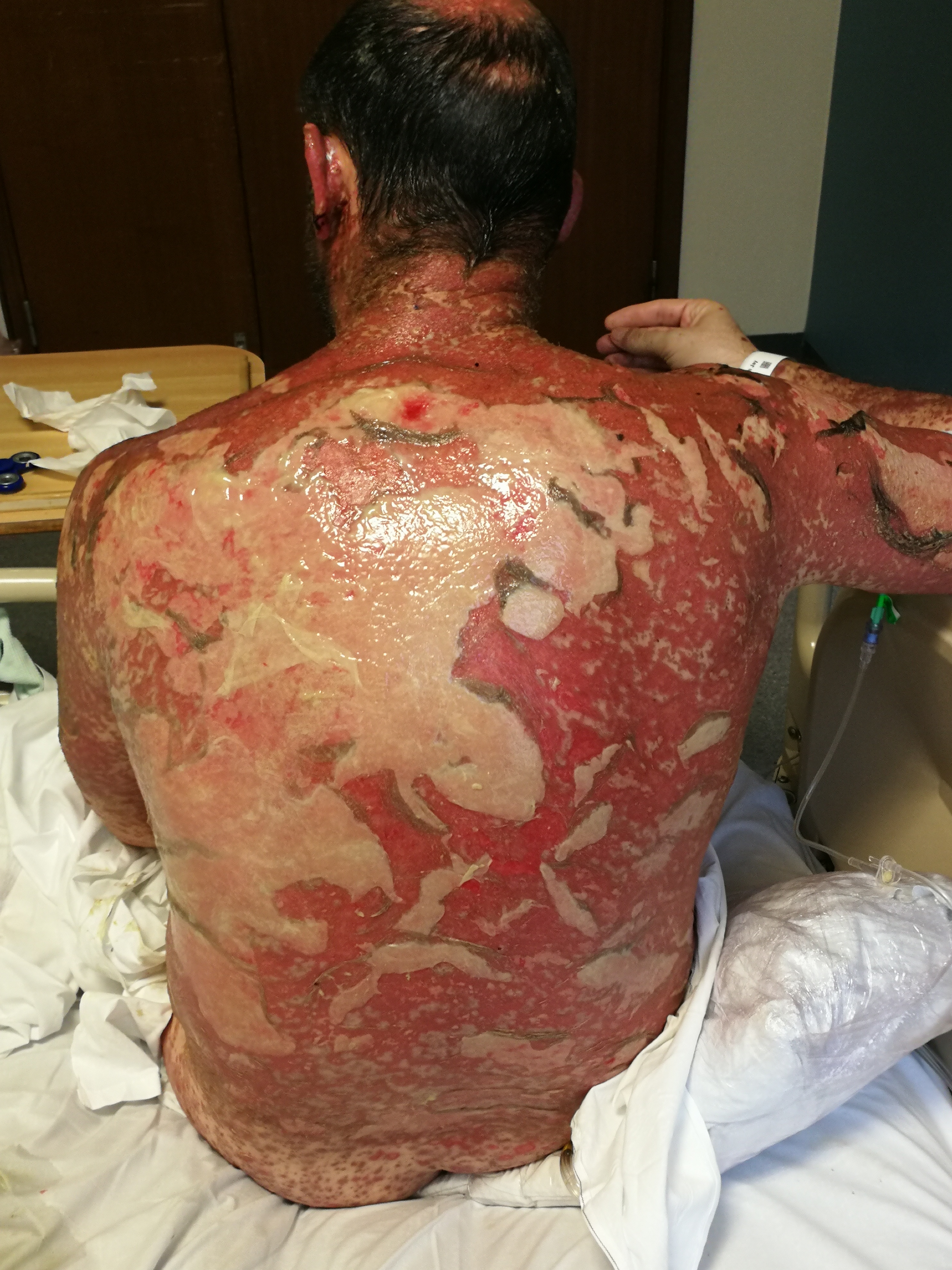
By <a href=”//commons.wikimedia.org/w/index.php?title=User:Dswierc&amp;action=edit&amp;redlink=1″ class=”new” title=”User:Dswierc (page does not exist)”>D Swierczek</a> – <span class=”int-own-work” lang=”en”>Own work</span>, CC BY-SA 4.0, Link

Erythema Gyratum Repens

  • Erythema gyratum repens is a rare paraneoplastic type of annular erythema with a ‘wood-grain’ appearance associated with malignancy. Furthermore, almost half of the patients with erythema gyratum repens have lung cancer and less commonly, oesophageal, breast, and stomach cancer.
  • The rash usually resolves once the malignancy has been removed with surgical resection.

References and Further Reading

Cite this article as: Leah Sarah Peer, Canada, "Erythema Types in Medicine – Rapid Review For Medical Students," in International Emergency Medicine Education Project, January 4, 2021, https://iem-student.org/2021/01/04/erythema-review-for-medical-students/, date accessed: October 1, 2023

Giant Hogweed Burns

Giant Hogweed…and the Lesions it Causes.

Summer is the time when outdoor work and leisure activities increase. It is also the season when plants like giant hogweed grow and bloom most. Direct contact with this beautiful umbrella plant, however, leads to serious skin lesions and skin burns[1] that often necessitates a visit to the emergency room.

Have you ever heard of the giant hogweed?

The giant hogweed is an extremely toxic exotic plant particularly present in the British popular culture. It is also mentioned in the 1971 studio album entitled “Nursery Cryme” by the English rock band Genesis where the eternal struggle between this exotic plant and the English authorities trying to eradicate it is (humorously) narrated. Historically this plant was rediscovered in 1895 by two Italian doctors: Emile Levier and Carlo Pietro Stefano Sommier, to which they gave the botanical name of Heracleum mantegazzanium, in honour of their friend and physician Paolo Mantegazza, the first popularizer of Darwin’s scientific theories in Italy.

How can you recognize giant hogweed?

Giant hogweed, also known as Heracleum mantegazzianum, is considered to be the largest and most beautiful umbrella plant in the world[2]; it can reach a height of over 3 meters[3]. It is recognizable due to the colour of its leaves, which are a bright light green colour tending towards yellow with deep lobes and segmentations. The trunk is very thick and robust, similar to that of artichoke, with dark red streaks and surrounded by spiky hairs. The diameter of the umbrella makes it the largest among the umbrella plants. 

The fruits have an ovoid appearance, and at the moment of flowering, their envelopes remain attached to the base of the umbrella and subsequently tend to wither. Giant hogweed blooms from early spring until late summer, especially in the vicinity of wetlands (streams or canal banks). These characteristics differentiate it from the garden angelica (Angelica archangelica) and the common hogweed (Heracleum sphondylium).

Who is most at risk of coming across giant hogweed?

Gardeners, trekkers, forest workers, and people who work outdoors in wooded or undergrowth areas where giant hogweed is present are most at risk of injury caused by giant hogweed.

How is giant hogweed sap toxic?

The sap of giant hogweed contains different photoactive agents called furocoumarins, of which the most predominant is 5-MOP (5-methoxypsoralen). Injuries are caused when these photoactive agents are exposed to and activated by UV-A rays present in sunlight. This gives rise to a toxic process in which the furan ring of the photoactive agent is cross-linked with the pyrimidine bases of DNA in the patient’s skin, thus causing an increase in oxidative stress leading to cell membrane damage and subsequent inflammation and oedema. The culmination of these processes leads to the development of phytophotodermatitis (PPD)[4].

Similar to other plants capable of phototoxicity, skin damage is dependent on certain factors[5] such as:

  • The concentration of the phototoxic agent
    • During summer months, levels of phototoxic agents are higher. Moreover, they are more concentrated in fruit, present in intermediate levels in the leaves, and are minimally present in the stem.
  • The thickness of the skin
    • Damage is more extensive and deeper in the skin that is thinner.
  • Sun exposure
    • When sun exposure is prolonged, there is greater photoactivation and therefore greater damage.
  • Skin moisture
    • Sweat or dew that may be present on the skin can accelerate the toxicity process.

What are its symptoms?

Symptoms of giant hogweed exposure usually involve an erythematous lesion[6] accompanied by extremely intense pain. If not treated early, erythematous lesions evolve into burns[7] with the appearance of one or more liquid-filled vesicles. Generally, patients may have fatigue and slight tachycardia, but vital signs and laboratory test may be normal.

During the history and physical examination, staphylococcal infection, allergic dermatitis, purpura, impetigo, and fungal infection must be considered in the differential diagnosis.

Therapy

Upon contact, the subject should immediately wash the red area abundantly, dry it, and cover it to avoid sun exposure. Other recommendations would include avoiding to take baths and showers and apply high protection sunscreen to the lesion.

If erythema appears, the use of sulfadiazine cream and analgesic anti-inflammatory drugs should be considered. Moreover, the use of ice to reduce inflammation can also be useful. In the event of an injury characterized by one or more blisters or loss of tissue, a conservative treatment[8] consisting of cleansing with antiseptic solution (i.e. Clorexidine) and bandaging with synthetic microporous membranes[9] have proven effective. In cases where the phototoxicity process has been prolonged enough to cause extensive and deep burns, skin grafting and surgical debridement are necessary.

Take-home message

In conclusion, the message for our patients who operate or live in areas where giant hogweed is present is to “look but absolutely don’t touch” this beautiful plant. Secondarily, it is also important to subsequently report its presence to public authorities; in fact, many countries or regions follow a specific eradication program for this plant which can prove dangerous for humans and animals.

Look but absolutely don't touch this beautiful plant.

References and Further Reading

[1] https://www.plymouthherald.co.uk/news/plymouth-news/man-suffers-horror-burns-hogweed-4216136

[2] Heracleum mantegazzianum (giant hogweed) – Invasive Species Compendium – https://www.cabi.org/isc/datasheet/26911

[3] Derraik JG. Heracleum mantegazzianum and Toxicodendron succedaneum: plants of human health significance in New Zea-land and the National Pest Plant Accord. N Z Med J 2007;120: U2657.

[4] Marcos LA, Kahler R. Phytophotodermatitis. Int J Infect Dis 2015;9(July (38)):7–8.

[5] Pira E, Romano C, Sulotto F, et al. Heracleum mantegazzianum growth phases and furocoumarin content. Contact Derm 1989; 21:300e3.

[6] J Emerg Nurs. 2006 Jun;32(3):246-8. A 43-year-old woman with painful, vesicular lesions from giant hogweed photodermatitis. Langley DM(1), Criddle LM.

[7] Baker BG, Bedford J, Kanitkar S. Keeping pace with the media; Giant Hogweed burns – A case series and comprehensive review. Burns. 2017 Aug;43(5):933-938. DOI: 10.1016/j.burns.2016.10.018.

[8] Chan JC, Sullivan PJ, O’Sullivan MJ, Eadie PA. Full thickness burn caused by exposure to giant hogweed: delayed presentation, histological features and surgical management. J Plast Reconstr Aesthet Surg. 2011;64(1):128-130. doi:10.1016/j.bjps.2010.03.030

[9] Pfurtscheller K, Trop M. Phototoxic plant burns: report of a case and review of topical wound treatment in children. Pediatr Dermatol 2014;31:e156–9.

Cite this article as: Francesco Adami, Italy, "Giant Hogweed Burns," in International Emergency Medicine Education Project, August 31, 2020, https://iem-student.org/2020/08/31/giant-hogweed-burns/, date accessed: October 1, 2023

A simple cellulitis of the foot?

a simple cellulitis of the foot?

Case Introduction

A 47 year old woman comes to a community ED complaining of pain and redness in her right foot developing quickly over two days. She denies any trauma and otherwise feels well. She is not sure, but may have had a “sore” near her toes that has already healed. Patient has diabetes but is normoglycemic. She has no prior history of cellulitis, joint infections or gout. There is no history of immunocompromise, including steroids, or any IV drug use. All vitals are within normal limits and review of systems is negative for fever, chills, respiratory or gastrointestinal symptoms.

On exam, there is generalized edema, erythema and tenderness, but no tenderness out of proportion, and no open sores or ulcerations. A sub-acute appearing callus is apparent on the plantar surface opposite fifth and fourth distal metatarsals. The ankle joint is tender but less so than the foot, and ranging it does not elicit more pain than at baseline. Distal sensation, pulses and toe motion are intact, though capillary refill is slightly delayed.

cellulitis - foot
cellulitis - foot 2

Initial Questions

Basic labs obtained are unremarkable and patient is receiving IV broad spectrum antibiotics, including MRSA coverage. Plain films are obtained, and there is some concern for small air pockets in the soft tissues.

cellulitis - xray 2
cellulitis - xray

A phone consultation with podiatry is obtained. A decision is made to take the patient to the OR on the same evening. No further imaging or diagnostic studies are advised.

Additional Questions

After the callus is taken off in the OR, large amount of frank pus is obtained that tracks all the way to the third metatarsal. A debridement is performed, and long term antibiotics with close follow up are needed. Overall impression was that while no necrotizing infection was found, any further delay would have risked a trans-metatarsal amputation (at the least).

Key Points

While we do not have room for a lengthy discussion on differentiating plain cellulitis from “other”, it is worthwhile to note several things:

Cite this article as: Anthony Rodigin, USA, "A simple cellulitis of the foot?," in International Emergency Medicine Education Project, February 7, 2020, https://iem-student.org/2020/02/07/educational-case-a-simple-cellulitis-of-the-foot/, date accessed: October 1, 2023

Further Reading

Anaphylaxis in a Nutshell

anaphylaxis in a nutshell

Anaphylaxis can be broadly defined as a severe, life-threatening, generalized or systemic hypersensitivity reaction. Literature suggests that anaphylaxis is not always easily recognized in the Emergency Department (ED). One study indicates around 50% of cases being misdiagnosed and up to 80% do not receive appropriate first-line treatment.

Triggers

The most commonly identified triggers of anaphylaxis include food, drugs and venom, but it is important to note that 30% of the cases can be idiopathic. Among drugs, muscle relaxants, antibiotics, NSAIDs and aspirin are the most commonly implicated.

Which patients are at an increased risk of anaphylaxis severity and mortality?

Extremes of age

Co-morbid conditions (asthma, COPD, cardiovascular disease)

Concurrent use of beta-blockers and ACE inhibitors

While the overall prognosis of anaphylaxis is good, the key to avoiding adverse outcomes is by prompt recognition and initiation of appropriate interventions. Below are key points to guide your management of anaphylaxis in the ED.

Recognizing Anaphylaxis in the ED

Anaphylaxis reactions vary significantly in duration and severity and a single set of criteria will not identify all anaphylactic reactions. The World Allergy Organization (WAO) has suggested the following criteria to help ED physicians be more consistent in their recognition of anaphylaxis.

Anaphylaxis is highly likely when any one of the following three criteria is fulfilled

1. Acute onset of an illness (minutes to several hours) with involvement of the skin, mucosal tissue, or both (eg, generalized urticaria, itching or flushing, swollen lips-tongue-uvula) AND AT LEAST ONE OF THE FOLLOWING

  • Respiratory compromise (eg, dyspnea, wheeze-bronchospasm, stridor, reduced PEF, hypoxemia)
  • Reduced blood pressure or associated symptoms of end-organ dysfunction (eg. hypotonia [collapse], syncope, incontinence) OR

2. Two or more of the following that occur rapidly after exposure to a likely allergen for that patient (minutes to several hours)

  • Involvement of the skin-mucosal tissue (eg, generalized urticaria, itch-flush, swollen lips-tongue-uvula)
  • Respiratory compromise (eg, dyspnea, wheeze-bronchospasm, stridor, reduced PEF, hypoxemia)
  • Reduced blood pressure or associated symptoms (eg, hypotonia [collapse], syncope, incontinence)
  • Persistent gastrointestinal symptoms (eg, crampy abdominal pain, vomiting) OR

3. Reduced blood pressure after exposure to known allergen for that patient (minutes to several hours)

  • Infants and children: low systolic blood pressure (age-specific) or greater than 30% decrease in systolic blood pressure
  • Adults: systolic blood pressure of less than 90 mm Hg or greater than 30% decrease from that person’s baseline

Management Algorithm of Anaphylaxis in the ED

Anaphylaxis algorithm
Anaphyaxis algorithm 2

Key Points in Management

References and Further Reading

Cite this article as: Neha Hudlikar, UAE, "Anaphylaxis in a Nutshell," in International Emergency Medicine Education Project, January 31, 2020, https://iem-student.org/2020/01/31/anaphylaxis-in-a-nutshell/, date accessed: October 1, 2023

Cellulitis – Clinical Image and Ultrasound

cellulitis

Case Presentation

A 45-years-old male with a week history of right leg swelling and redness presented to the ED. He has type II DM and hypertension. He denies fever; however, complaints about burning pain over the skin. Vitals were 156/98 mmHg blood pressure, 98 beats per minute heart rate, 16 respiration per minute, 36.7 degrees Celsius temperature and 98% oxygen saturation in room air. Physical exam revealed erythema over the right medial lower leg and calf area (images). Minimally painful with palpation. The area was hot compared to the left leg. Other examination findings were unremarkable.

Cellulitis 2

Cellulitis 1

Patients with red, swollen, painful leg may have very severe problems such as necrotizing fasciitis (infection involving muscular fascia) or infections involving muscles with or without gangrene. The patients having these infections are generally ill-looking, severely painful, and may have subcutaneous crepitations. Therefore, we should be aware of these red flags. This patient has no sign of crepitations, systemic illness, or severe pain.

Lipodermatosclerosis is chronic erythema. Patients show exacerbations because of vascular insufficiency (venous). It can be bilateral or unilateral. One of the discriminative findings from cellulitis is temperature over the lesion. Lipodermatosclerosis is not hot. In the case, the palpation showed warm skin compared to the left side.

Erysipelas is superficial and its’ borders are very sharp. The lesion is fluffy compared to the skin around the lesion. In the case, some areas of the skin were found a little bit raised compared to surrounding structures. However, its’ borders were not well-demarcated.

Other differentials are burns, contact dermatitis, urticaria, etc.

Bedside ultrasound imaging can help to identify cellulitis, abscess, foreign body, fracture, etc. Cobblestone finding is a typical finding for cellulitis.

Bedside ultrasound imaging was performed with Butterfly iQ with soft tissue settings. Cobblestone finding was found in the erythematous areas. This is a nonspecific finding and can be seen many different soft tissue infections. There were no gas/air artifacts (necrotizing fasciitis) or obvious abscess formation. However, there was a minimal fluid accumulation, which creates a suspicion of an abscess. In the case, there was no air artifact. However, x-rays can also help to show air accumulation in soft tissues.

An Example for Necrotizing Fasciitis

The ultrasound investigation in this video shows the air (white) artifacts in the soft tissue.

X-ray Image Showing Subcutaneous Air in Necrotizing Fasciitis

Case courtesy of Dr Matt Skalski, Radiopaedia.org. From the case rID: 25026

For mild uncomplicated patients – dicloxacillin, amoxicillin, and cephalexin are common choices.

If the patient has a penicillin allergy – clindamycin or a macrolide (clarithromycin or azithromycin) can be used.

Fluoroquinolones should be reserved for gram-negative organisms’ sensitivity defined by culture results because of their additional toxicity risks.

For more antibiotic options and explanations, please visit – here

The patients with co-morbidities compromising immune response, periorbital or perianal locations, unable to tolerate oral medication, deep infections should be admitted.

References and Further Reading

  • Loyer EM, DuBrow RA, David CL, Coan JD, Eftekhari F. Imaging of superficial soft-tissue infections: sonographic findings in cases of cellulitis and abscess. AJR Am J Roentgenol. 1996 Jan;166(1):149-52. PubMed PMID: 8571865.
  • Shyy W, Knight RS, Goldstein R, Isaacs ED, Teismann NA. Sonographic Findings in Necrotizing Fasciitis: Two Ends of the Spectrum. J Ultrasound Med. 2016 Oct;35(10):2273-7. doi: 10.7863/ultra.15.12068. Epub 2016 Aug 31. PubMed PMID: 27582527.
Cite this article as: Arif Alper Cevik, "Cellulitis – Clinical Image and Ultrasound," in International Emergency Medicine Education Project, December 2, 2019, https://iem-student.org/2019/12/02/cellulitis-clinical-image-and-ultrasound/, date accessed: October 1, 2023

Clinical Image: rhabdomyosarcoma?

862.1 - rhabdomyosarcoma 1

A 35-year-old male with a seven-month history of right supraclavicular mass. No compressive symptoms. Clinical and Xray interpretation was soft tissue rhabdomyosarcoma.

862.2 - rhabdomyosarcoma 2
862.3 - rhabdomyosarcoma 3

Rhabdomyosarcoma is one of the aggressive and malignant cancers of skeletal (striated) muscle cells. The cases are mostly young, particularly below age 18. It may arise from al body regions. However, head and neck, urinary and reproductive system, extremities are common locations.

Do you need more free clinical images or videos for your exams or presentations? Please visit iEM clinical image and video archive in Flickr and YouTube!

Cite this article as: Masuma Ali Gulamhussein, "Clinical Image: rhabdomyosarcoma?," in International Emergency Medicine Education Project, July 8, 2019, https://iem-student.org/2019/07/08/clinical-image-rhabdomyosarcoma/, date accessed: October 1, 2023

An 8-year-old male with erythematous lesions

713 - tinea capitis and corporis - dog in the house

An 8-year-old male presented to the ED with erythematous papules appeared one week ago. He has one lesion in the scalp and another in his left lower limb over the calf muscle. The patient is complaining of pain and itchiness over the lesions. His brother was admitted the day before for a similar complaint. Both were exposed to a pet dog at home.

What is your diagnosis? – the answer is here.