Dermatological emergencies : Stevens-Johnson Syndrome

International Emergency Medicine Education Project
We promote emergency medicine and provide free, reusable education resources for medical students and educators
Category 3 catches you by surprise when it makes it an entry in the ED and serves as a reminder of why it is essential always to know something about everything. Stevens-Johnson Syndrome was one of those for me. Although rare, dermatological emergencies are essential to spot and can be life-threatening if left untreated.
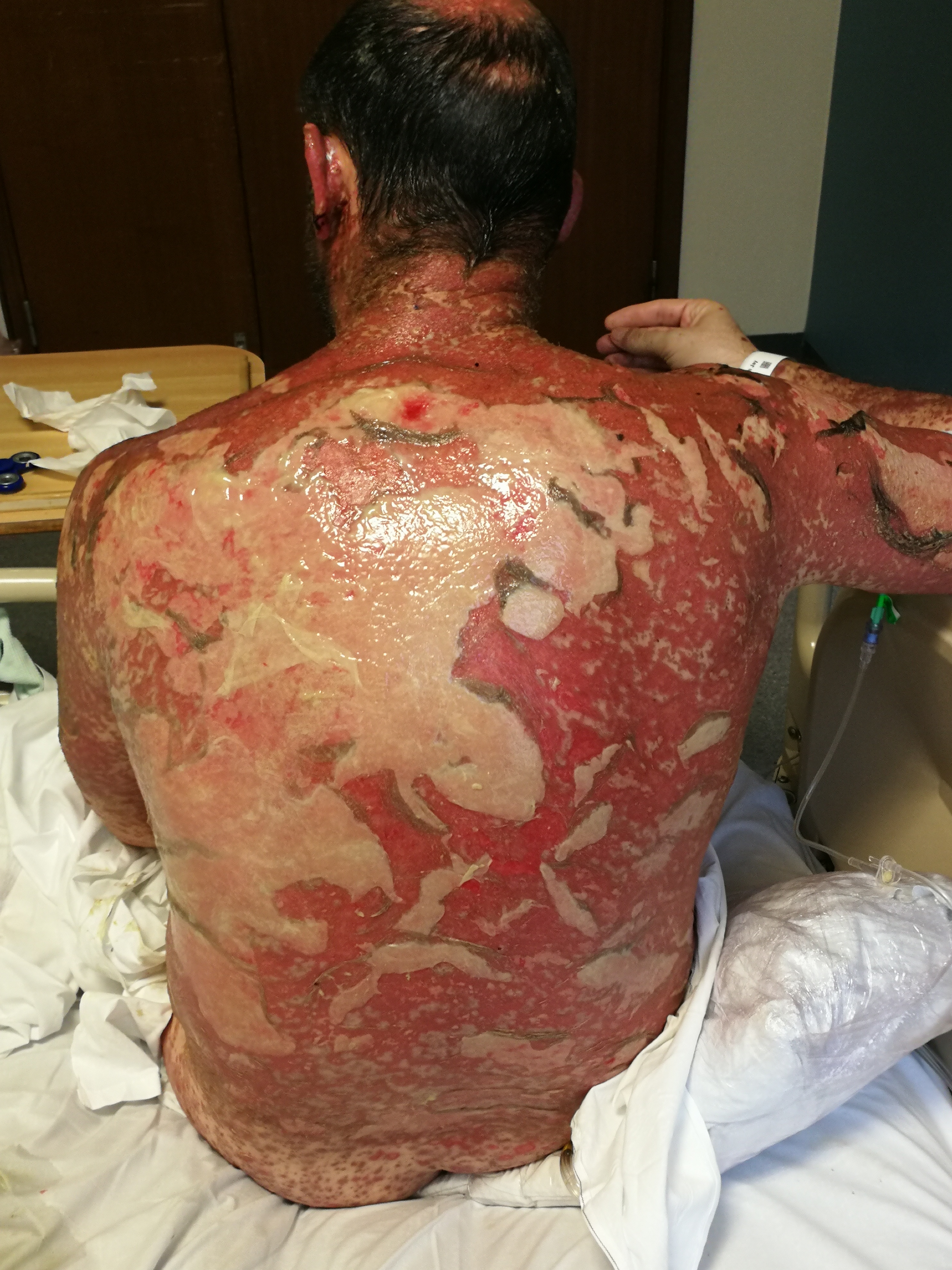
Stevens-Johnsons Syndrome is a rare type 4 hypersensitivity reaction which affects <10% of body surface area. It is described as a sheet-like skin loss and ulceration (separation of the epidermis from the dermis).
Toxic epidermal necrosis and Stevens-Johnsons Syndrome can be mixed. However, distinguishing between both disease can be done by looking at % of body surface area involvement.
Pathophysiology is not clearly known; however, some studies show it is due to T cells’ cytotoxic mechanism and altered drug metabolism.
The most common cause of Stevens-Johnsons Syndrome is medications. Examples are allopurinol, anticonvulsants, sulfonamide, antiviral drugs, NSAIDs, salicylates, sertraline and imidazole.
As one of the commonest cause is drug-induced, it is a vital part of history taking. Ask direct and indirect questions regarding drug intake, any new (started within 8 weeks) or old medications and previous reactions if any.
Other causes are malignancy and infections (Mycoplasma pneumonia, Cytomegalovirus infections, Herpesvirus, Hep A).
The disease is more common in women and immunocompromised patients (HIV, SLE)
Diseases with a similar presentation – in children, staphylococcal scalded skin syndrome can be suspected as it has a similar presentation and can be differentiated with the help of a skin biopsy.
Clinical awareness and suspicion is the cornerstone step for diagnosis. Skin Biopsy shows subepidermal bullae, epidermal necrosis, perivascular lymphocytic infiltration, which help for definitive diagnosis.
Adequate fluid resuscitation, pain management and monitoring of electrolytes and vital signs, basic supportive or resuscitative actions are essential, as with any emergency management.
The next step is admitting the patient to the burn-unit or ICU, arranging an urgent referral to dermatology and stopping any offending medications. If any eye symptoms are present, an ophthalmology referral is required.
Wound management is essential- debridement, ointments, topical antibiotics are commonly used to prevent bacterial infections and ease the symptoms.
Prognosis of a patient with Stevens-Johnson Syndrome is assesed by the SCORTEN Mortality Assesment Tool. Each item equal to one point and it is used within the 24 hours of admission.
• Age >/= 40 years (OR 2.7)
• Heart Rate >/= 120 beats per minute (OR 2.7)
• Cancer/Hematologic malignancy (OR 4.4)
• Body surface area on day 1; >10% (OR2.9)
• Serum urea level (BUN) >28mg/dL (>10mmol/L) (OR 2.5)
• Serum bicarbonate <20mmol/L (OR 4.3)
• Serum glucose > 252mg/dL (>14mmol/L) (OR5.3)
Predicted mortality based on the above total:

One of the most convenient ways of learning and remembering the main components of disease and identifying a medical condition on an exam are Triads, and medical students/interns/residents swear by them.
Be it a question during rounds, a multiple-choice exam question to be solved, or even in medical practice, the famous triads help physicians recall important characteristics and clinical features of a disease or treatment in an instant.
Since exam season is here, this could serve as a rapid review to recall the most common medical conditions.
While there are a vast number of triads/pentads available online, I have listed the most important (high-yy) ones that every student would be asked about at least once in the duration of their course.
1) Lethal Triad also known as The Trauma Triad of Death
Hypothermia + Coagulopathy + Metabolic Acidosis
2) Beck’s Triad of Cardiac Tamponade
Muffled heart sounds + Distended neck veins + Hypotension
3) Virchow’s Triad – Venous Thrombosis
Hypercoagulability + stasis + endothelial damage
4) Charcot’s Triad – Ascending Cholangitis
Fever with rigors + Right upper quadrant pain + Jaundice
5) Cushing’s Triad – Raised Intracranial Pressure
Bradycardia + Irregular respiration + Hypertension
6) Triad of Ruptured Abdominal Aortic Aneurysm
Severe Abdominal/Back Pain + Hypotension + Pulsatile Abdominal mass
7) Reactive Arthritis
Can’t See (Conjunctivitis) + Can’t Pee (Urethritis) + Can’t Climb a Tree (Arthritis)
8) Triad of Opioid Overdose
Pinpoint pupils + Respiratory Depression + CNS Depression
9) Hakims Triad – Normal Pressure Hydrocephalus
Gait Disturbance + Dementia + Urinary Incontinence
10) Horner’s Syndrome Triad
Ptosis + Miosis + Anydrosis
11) Mackler’s Triad – Oesophageal Perforation (Boerhaave Syndrome)
Vomiting + Lower Thoracic Pain + Subcutaneous Emphysema
12) Pheochromocytoma
Palpitations + Headache + Perspiration (Diaphoresis)
13) Leriche Syndrome
Buttock claudication + Impotence + Symmetrical Atrophy of bilateral lower extremities
14) Rigler’s Triad – Gallstone ileus
Gallstones + Pneumobilia + Small bowel obstruction
15) Whipple’s Triad – Insulinoma
Hypoglycemic attack + Low glucose + Resolving of the attack on glucose administration
16) Meniere’s Disease
Tinnitus + Vertigo + Hearing loss
17) Wernicke’s Encephalopathy- Thiamine Deficiency
Confusion + Ophthalmoplegia + Ataxia
18) Unhappy Triad – Knee Injury
Injury to Anterior Cruciate Ligament + Medial collateral ligament + Medial or Lateral Meniscus
19) Henoch Schonlein Purpura
Purpura + Abdominal pain + Joint pain
20) Meigs Syndrome
Benign ovarian tumor + pleural effusion + ascites
21) Felty’s Syndrome
Rheumatoid Arthritis + Splenomegaly + Neutropenia
22) Cauda Equina Syndrome
Low back pain + Bowel/Bladder Dysfunction + Saddle Anesthesia
23) Meningitis
Fever + Headache + Neck Stiffness
24) Wolf Parkinson White Syndrome
Delta Waves + Short PR Interval + Wide QRS Complex
25) Neurogenic Shock
Bradycardia + Hypotension + Hypothermia

Starting the Emergency Medicine (EM) Clerkship is one of the most exciting times of any medical student’s life, regardless of whichever specialty they plan on specializing in because EM has something for everyone. It is like solving all those questions that begin with ‘A patient presents to the Emergency Department with…’ but in reality, at a faster pace and with more tricky situations. This can make students feel overwhelmed, as they find themselves juggling between books and resources as to which one to follow or which topics to learn, and I am here for just that! To share the approach that helps many students get the hang of EM and make the most of their time in one of the best learning environments of any hospital.
The basic approach would be first to jot down all the problems you can think of.
Here is a list to help you get started: Core EM Clerkship Topics
There are problems that you may be heard a lot such as Chest Pain, Heart Failure, Shock (and it’s types), Acute Coronary Syndrome, Sepsis, Pulmonary edema, Respiratory Failure, Coma, Stroke, Hypoglycemia, Subarachnoid Hemorrhage, Fractures, Head Trauma, Status Epilepticus, Diabetic Ketoacidosis, and Anaphylaxis.
As every doctor you meet will always say, common is common, so always focus on things that you have heard and seen most about, read about them, make notes on their clinical features, differentials, investigations and management. Most importantly, do not forget to read about the ABCDE approach in every critically ill and trauma patient.
Know what to ask and when to ask. Patients in the ED are not in their most comfortable composure, so try to practice and frame questions that provide you with just enough information to make a diagnosis in the least possible time.
The same goes for examination, never forget the basics of examination and their importance. Practice examination as much as you can and you will automatically see it come to you naturally at a faster pace. Also, do not forget focused history and physical examination is a cornerstone of EM practice and saves a lot of time.
Where investigations can help you exclude a differential, 80% of your diagnosis will be built from what you ask, what you see and what you feel. Keep in mind that if you are not thinking or looking for something, you will not see and find it. So, be suspicious of life, organ and limb-threatening problems.
ABG, Intubation, Central Lines, FAST Scan, Suturing, Catheter and Cannula placement are some of them. As a medical student, you will probably not be required to perform any, but it is good to have an idea about the procedures when you see them. If you can practice, then that is even better, ask a resident or intern to show you how and you can have a go yourself under their supervision! Remember, “see one, do one, teach one.”
Youtube is an asset when it comes to medical education, make good use of it. There are also plenty of videos on the iEM website that you can watch and learn from.
Google is your best friend for this! You have the list of common conditions, all you need to do is a google search on the most common ECG findings and x-rays in medical emergencies and you will be good to go. You can also always learn these from the doctors around you in the ED, as the more you see and try to interpret, the better you get at differentiating the normal from abnormal.
Before the rotation, read a review book, recall your basic knowledge from internal medicine/family medicine and surgery because EM almost covers all of the acute problems of those fields. Moreover, do not forget, EM is an independent specialty and has its’ own textbooks.
iEM Clerkship book is a very good source to get started with! Download Now! – iEM Book (iBook and pdf)
If you are the kind, who likes solving questions, the Pretest Emergency Medicine is a great source.
During the rotation – Learning what you see is the best way to keep things in your long term memory. After your shift ends, and you go home, get some rest, recall the cases of the day and read about them on Up to Date/ Medscape or any resource that you prefer, this will help you relate what you saw with what you are reading and will help you recall it better later on.
These are just a few tips to help in making the most of your EM rotation. Remember to study hard, but also practice, brush up on your communication skills, talk to patients, be there for them. The EM Clerkship prepares you for life as a doctor, as you practice every aspect of medicine during this time and learn to answer questions about acute medical problems and their severity when asked by those around you.

When I was rotating on surgery as a medical student, our attending once asked of our small group what may be concerning in the differential for right upper quadrant abdominal pain. A very eager and a somewhat brash student blurted immediately: “Echinococcal cyst!” The attending replied, “Well, that’s true, but if Echinococcal cyst is the first thing you think of as a surgical consultant, you’re crazy!”
On the other hand, take a practicing internal medicine physician like my Dad. He formulates his differentials with a very different strategy, which is: what is the most likely? A chronic cough is bronchitis (even with hemoptysis), pneumonia, GERD or postnasal drip. Shoulder pain is, of course, a sprain, bursitis, or some referred cervical impingement. And so on.
Unfortunately, neither hunting for zebras (an unofficial US name for exciting but rare diagnoses) nor settling for the most common works for emergency medicine. In fact, that is how true diagnoses may get missed and patients may start dying.
The EM differential diagnosis is a pyramid tipped on its head. It is therefore different from how differentials are approached by many other specialties.
In EM, we first have to think of and rule out the most severe or threatening pathology. That’s a given. But our choices have to come from among the common killers, not Martian viruses or unheard of tumors from a medical encyclopedia.
Amoebic meningitis is exciting to encounter in your practice. But guess what? Your patient won’t have it. At the same time, for EM physicians things like pulmonary embolus, aneurysm of the abdominal aorta, subarachnoid hemorrhage and necrotizing fasciitis are everyday icons on our cognitive desktops. While less common than a common cold, these things are by no means rare.
In EM, one can rest assured that common pathology will present atypically and not quite like the textbook.
It may take years of practice to be able to persuade someone that you have done due diligence and your professional duty by excluding a whole lot of deadly things, while the exact diagnosis still remains elusive.
We are confident, but also afraid. We have to think of the worst yet possible scenario for any complaint, yet of course anticipate that the actual diagnosis will hopefully be something less severe and quite common – like a migraine. After all, after most CT scans and lumbar punctures, it is not a subarachnoid hemorrhage.
In EM, we are in this perpetual struggle with having to be professionally pessimistic and paranoid on the one hand, yet emotionally supportive and reassuring for the patient on the other. I always teach my students, even nursing trainees, that no one should be leaving an emergency department more scared or anxious than when they came in.
Your job as a rotating trainee in EM is to understand and learn this exact interplay.
For your attending, but more importantly for yourself and your patients, you have to be as concerned with sepsis from PID on a 16-year-old young woman with fever and abdominal pain as you are with appendicitis. The 86 year old grandmother with Afib but on no anti-coagulation, because she falls a lot is not just TIA or CVA prone. Her embolic clots may just as well be traveling downstream, causing that intermittent or out of proportion abdominal pain called mesenteric ischemia – for which you do not have a good lab test or imaging, by the way.
think of several real worst-case scenarios that are not zebras. Can something horrible yet by no means unheard of be presenting atypically? What steps can you take to prove or disprove it?
What else could be going on? Like a stack of dominoes: what happened first, what happened next?
while carefully and patiently pursuing your own professional agenda.
don’t forget to discuss worrisome signs for which to return. What if you’re still wrong?

Welcome to iEM Weekly Feed!
With this feed, you do not miss anything. You will find all published blog posts during this week. Click the “title” or “read more” to open each page you interested in.
by Elif Dilek Cakal Case Presentation A 45-year-old female with no prior medical history presented to the emergency department (ED) with three days of constant shortness
Do you recognize these findings in the US and CT scan? https://youtu.be/Dh-Q23xdqeEhttps://youtu.be/lhops90o6_g Pulmonary Embolism by Elif Dilek Cakal from Turkey. Read Listen
by Elif Dilek Cakal Case Presentation An 85-year-old woman, with a history of congestive heart failure, presented with right leg pain and swelling of 2
A New Chapter Is Just Uploaded To The Website! An 85-year-old woman, with a history of congestive heart failure, presented with right leg pain and
Ibrahim Sarbay Turkey I will never forget the time that I acted as a “medical doctor” at the 1st year-end show of the elementary school.
In case you didn’t encounter a child fallen from a bicycle today! Read “Multiple Trauma” Chapter Listen “Multiple Trauma” Chapter iEM Education Project Team uploads
Core EM clerkship topics recommended by SAEM are ready for students. Feel free to read or listen. And, do not forget to share with your
In case you didn’t encounter flank pain today! iEM Education Project Team uploads many clinical picture and videos to the Flickr and YouTube. These images
Uploading Image and Video Archive More than 350 images and videos are uploaded into iEM Flickr channel. Thousands to come… Uploading Image and Video Archive
You are the emergency doc working in a rural ED. It is the Saturday night at 23:25 and you have three patients with chest pain.
In case you didn’t encounter a patient with testicular pain today! iEM Education Project Team uploads many clinical picture and videos to the Flickr and
Uploading Audio Chapters 68% 28 chapters are ready to listen and download, 100 to go…
by Walid Hammad – USA Case Presentation An ambulance crew rushes into your emergency department (ED) with a 56-year-old man. He is severely short of
A New Chapter Is Just Uploaded To The Website! An ambulance crew rushes into your emergency department with a 56-year-old man. He is severely short
In case you didn’t encounter a kid with wrist pain today! Pediatric fractures affecting growth plate are classified with Salter-Harris classification. It is from I

by Maryam AlBadwawi Introduction Shock, in simple terms, is a reduced circulatory blood flow state within the body. The inadequate circulation deprives the tissues of its
by Asaad S Shujaa Introduction Chest pain is one of the most common symptoms presented in the emergency department (ED), and it is worrisome because
by Moira Carrol, Gurpreet Mudan, and Suzanne Bentley Case Presentation A 61-year-old man with a history of liver cirrhosis secondary to chronic EtOH abuse presents to
by Matevz Privsek and Gregor Prosen Introduction Headache is a subjective feeling of pain, crushing, squeezing or stabbing anywhere in the head. They are typically
by Ebru Unal Akoglu Case Presentation A 40-year-old female with a history of diabetes mellitus presents with a complaint of 6 days cough and muscle
by Pia Jerot and Gregor Prosen Case Presentation A 28-year old male was a restrained driver in a head-on motor vehicle collision. He was entrapped and
by Harajeshwar Kohli and Ziad Kazzi Case An 18-year-old, previously healthy female, presents to the Emergency Department with nausea, vomiting, and tremors. She states 45
by Abdel Noureldin and Falak Sayed Quick link to Spanish Version Introduction A 23-year-old female was brought into the emergency department. Her frantic family members
by Murat Cetin, Begum Oktem, Mustafa Emin Canakci Case Presentation An 80-year-old female presents to the emergency department with a tendency to sleep (altered mental
by Shaza Karrar Case Presentation A 39-year-old female presented to the emergency department (ED) complaining of right-lower-quadrant (RLQ) pain; pain duration was for 1-day, associated

This chapter describes how and why important the emergency medicine clerkship is. Although it aims to reach medical student/interns, there are many lessons to learn for us, educators.
The Emergency Medicine (EM) clerkship typically takes place during the fourth year of medical school. However, some programs may have an optional elective during the third year. Whether or not you plan to specialize in Emergency Medicine, the rotation is an important aspect of your medical education. The emergency room is a unique learning environment which is different than any other setting in the hospital. It provides clinical opportunities that are largely unavailable in other clerkships and rotations. During residency, many specialties will also spend a significant amount of time in the Emergency Department (ED). This may be within a structured EM rotation, or while admitting or seeing patients for a certain medical or surgical service. Therefore, it is important to gain an understanding of the flow of the ED as well as the particular thought process that must be employed with emergency department patients…


Continue reading “Core EM Clerkship Topics” →


